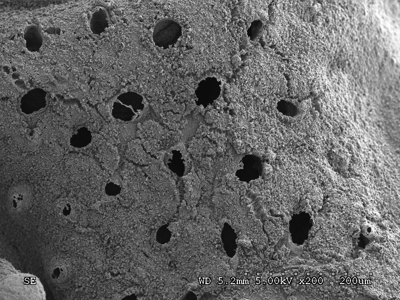
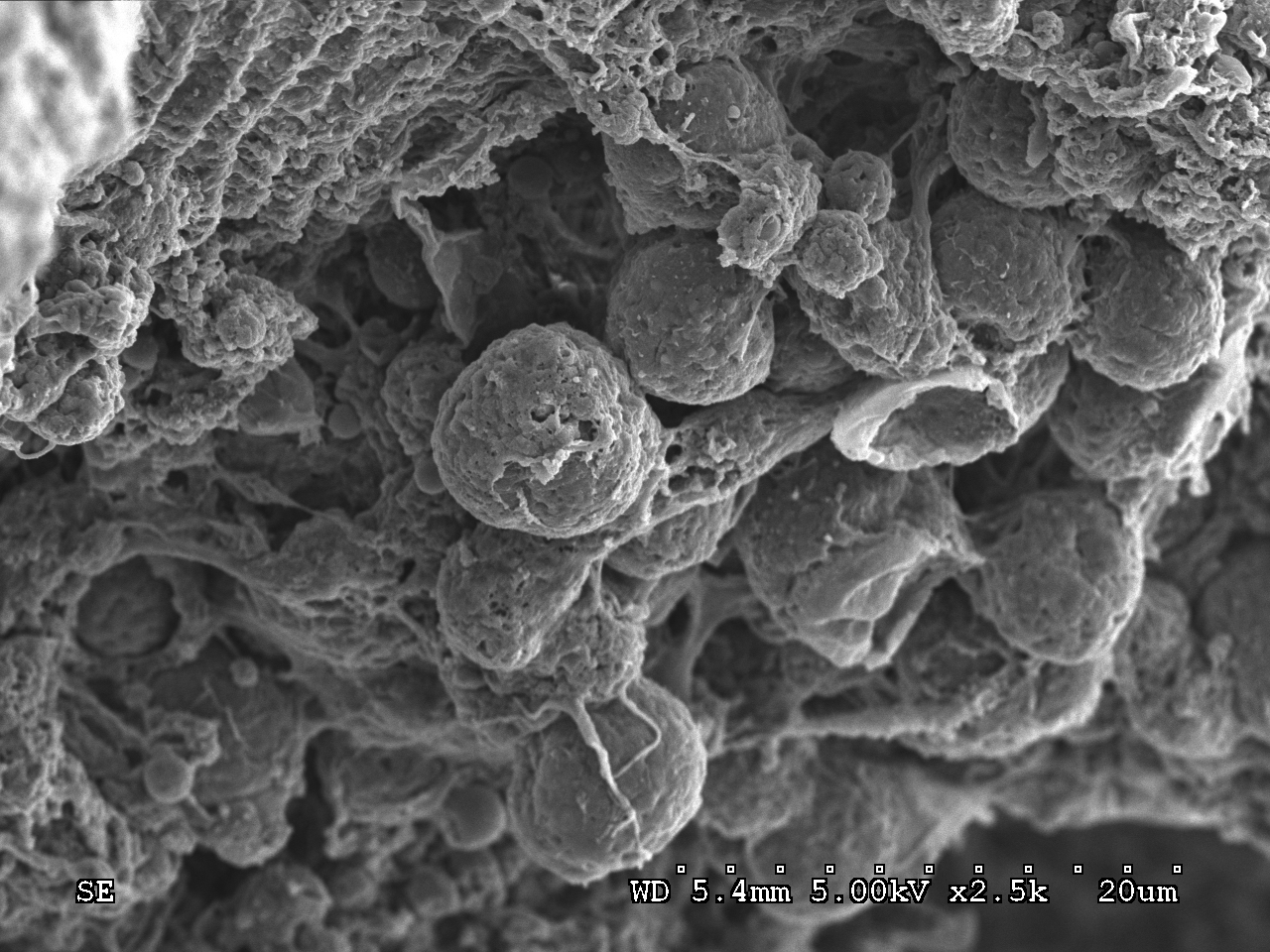
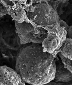
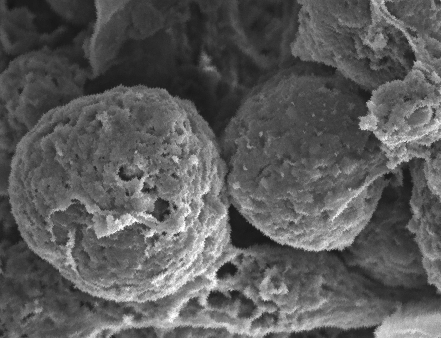
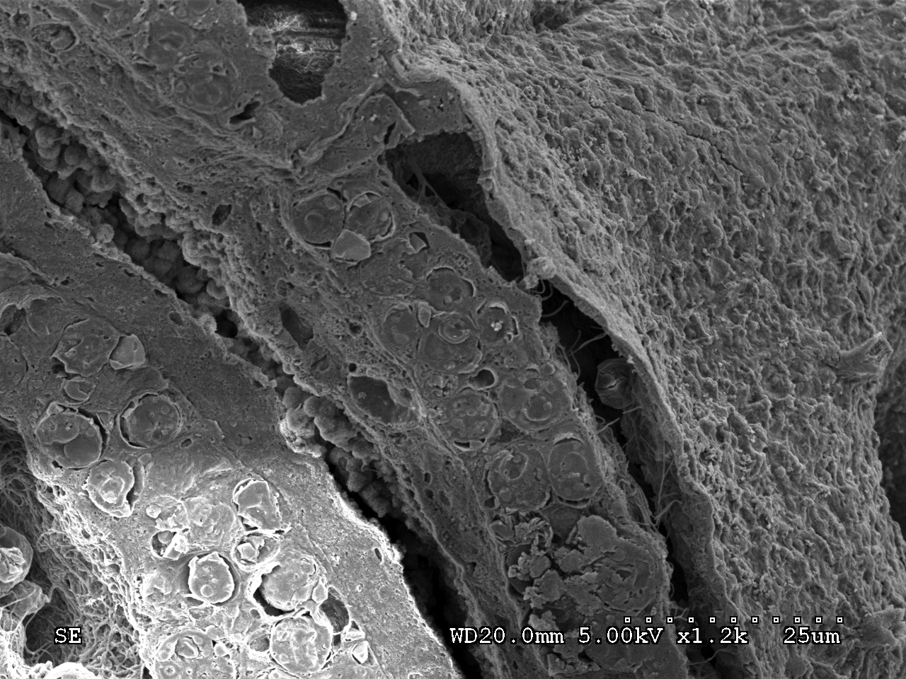
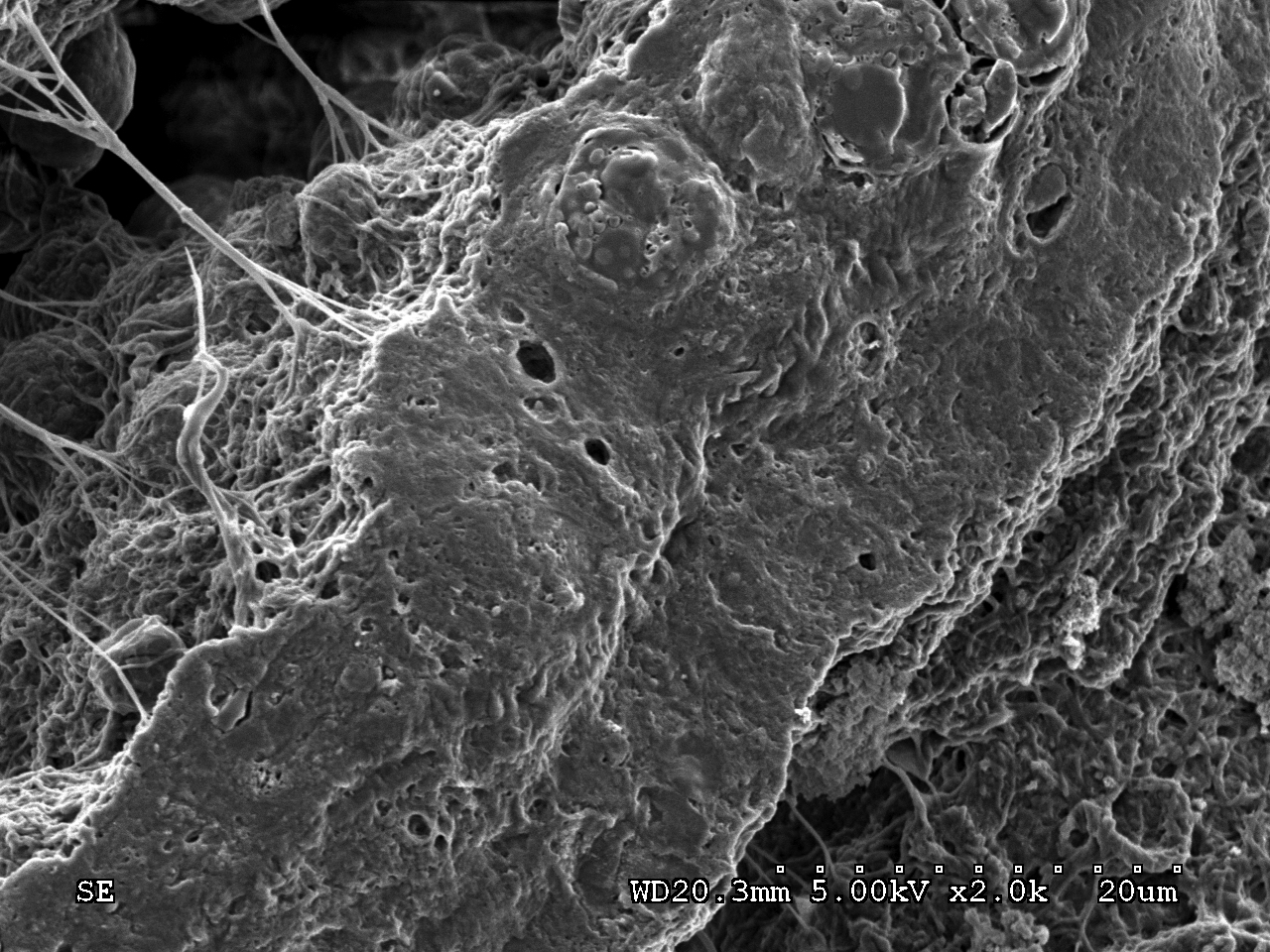
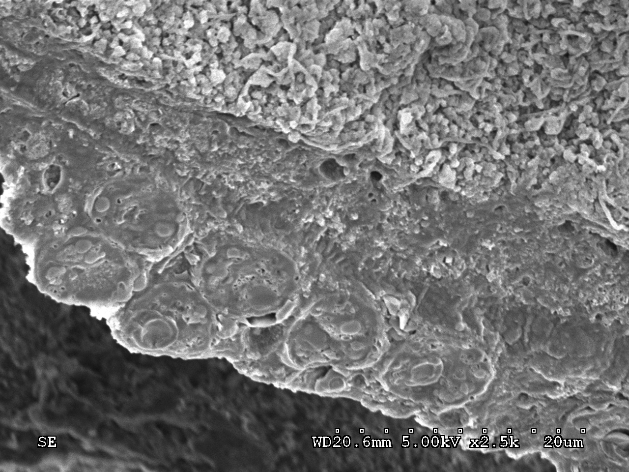
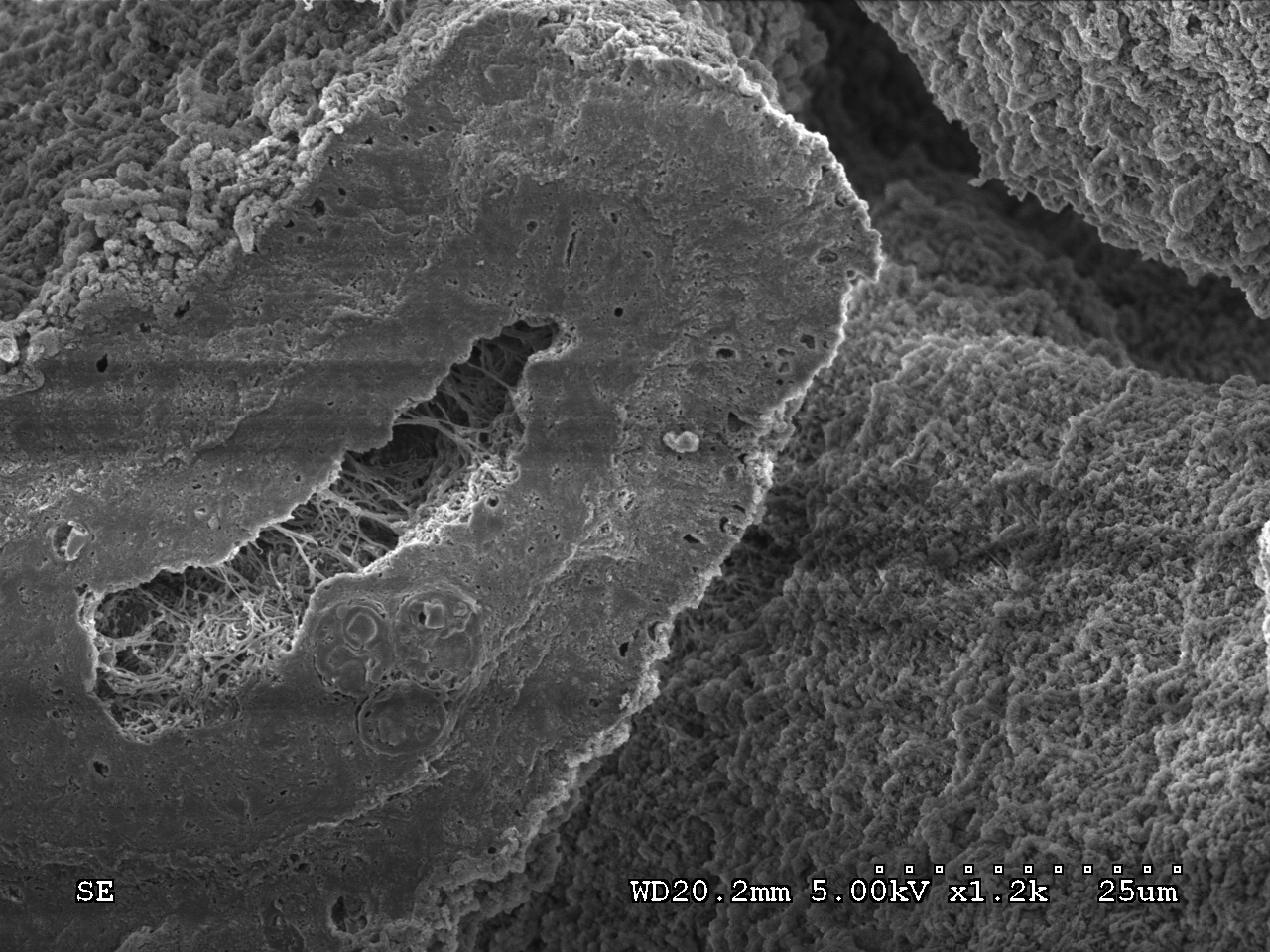
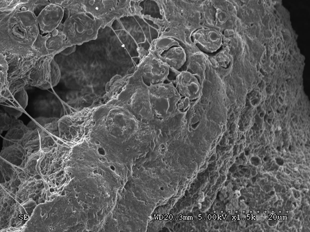
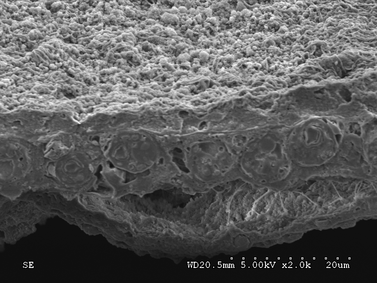
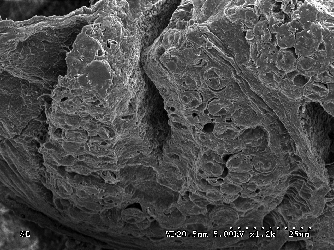
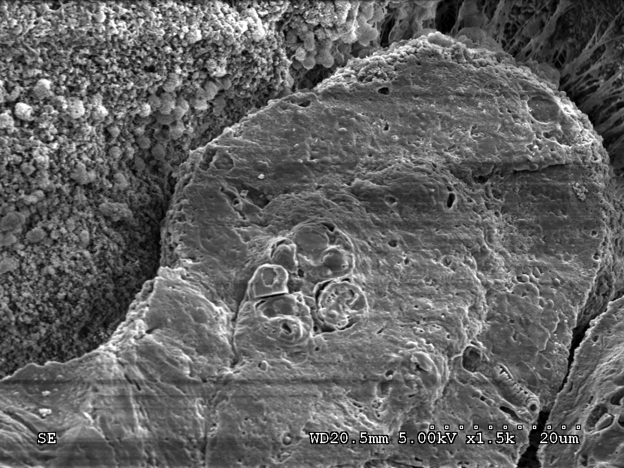
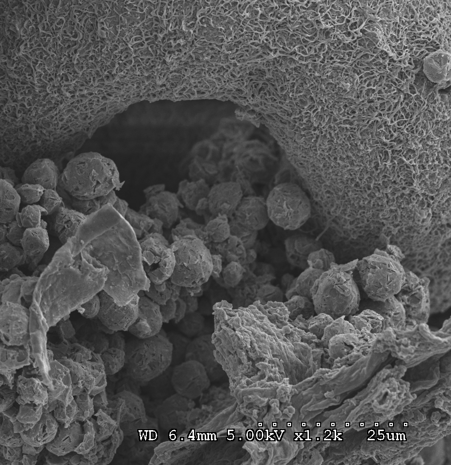
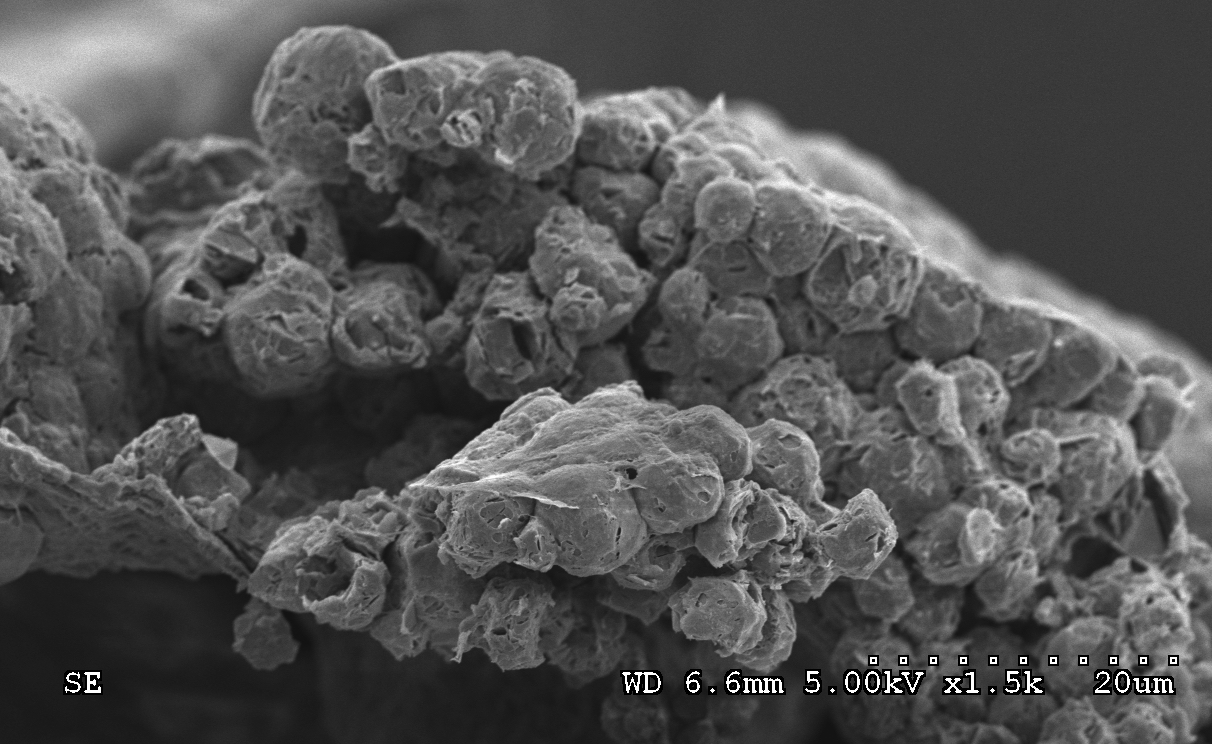
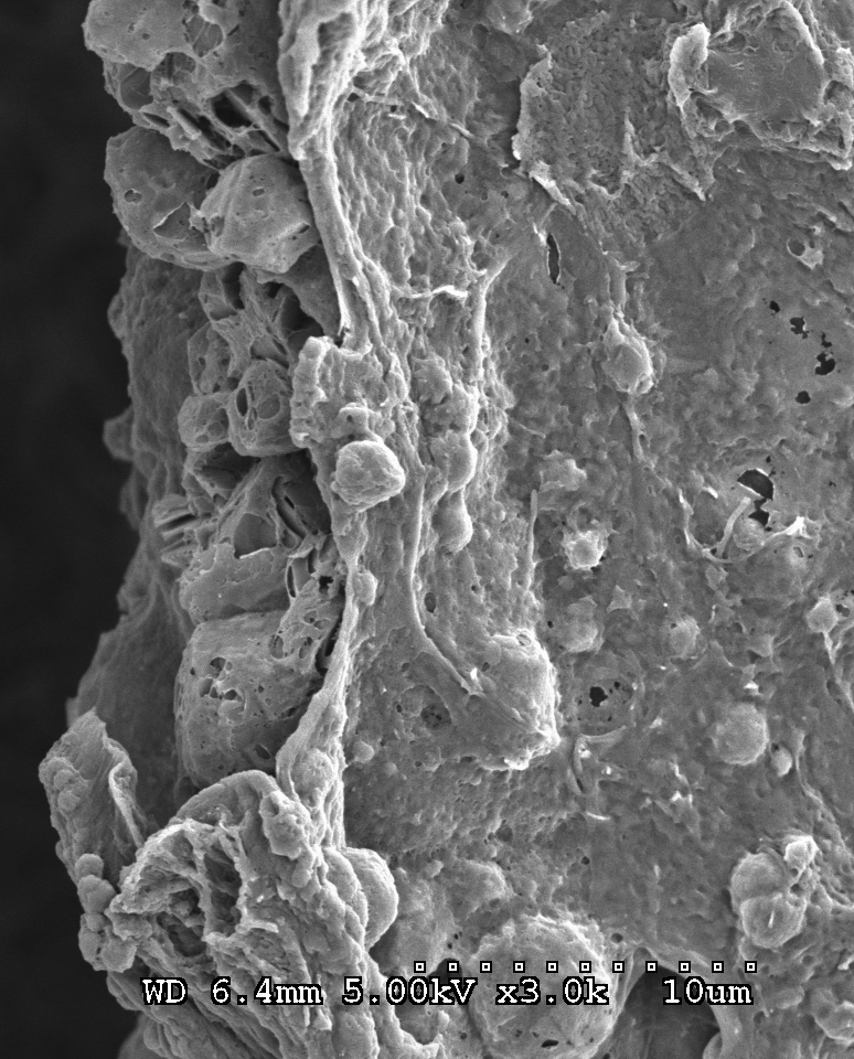
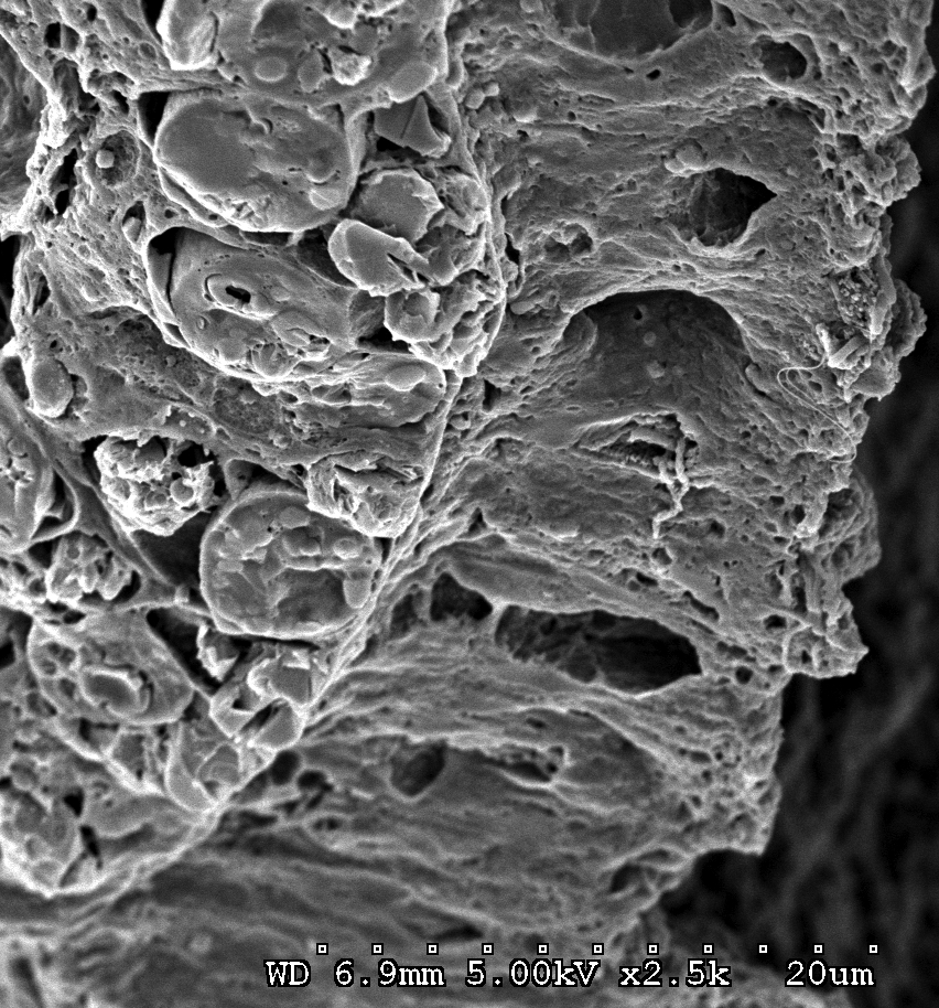
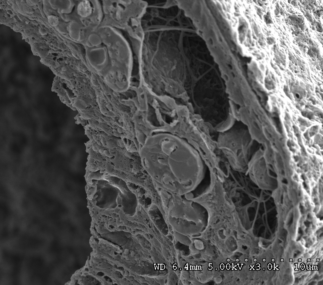
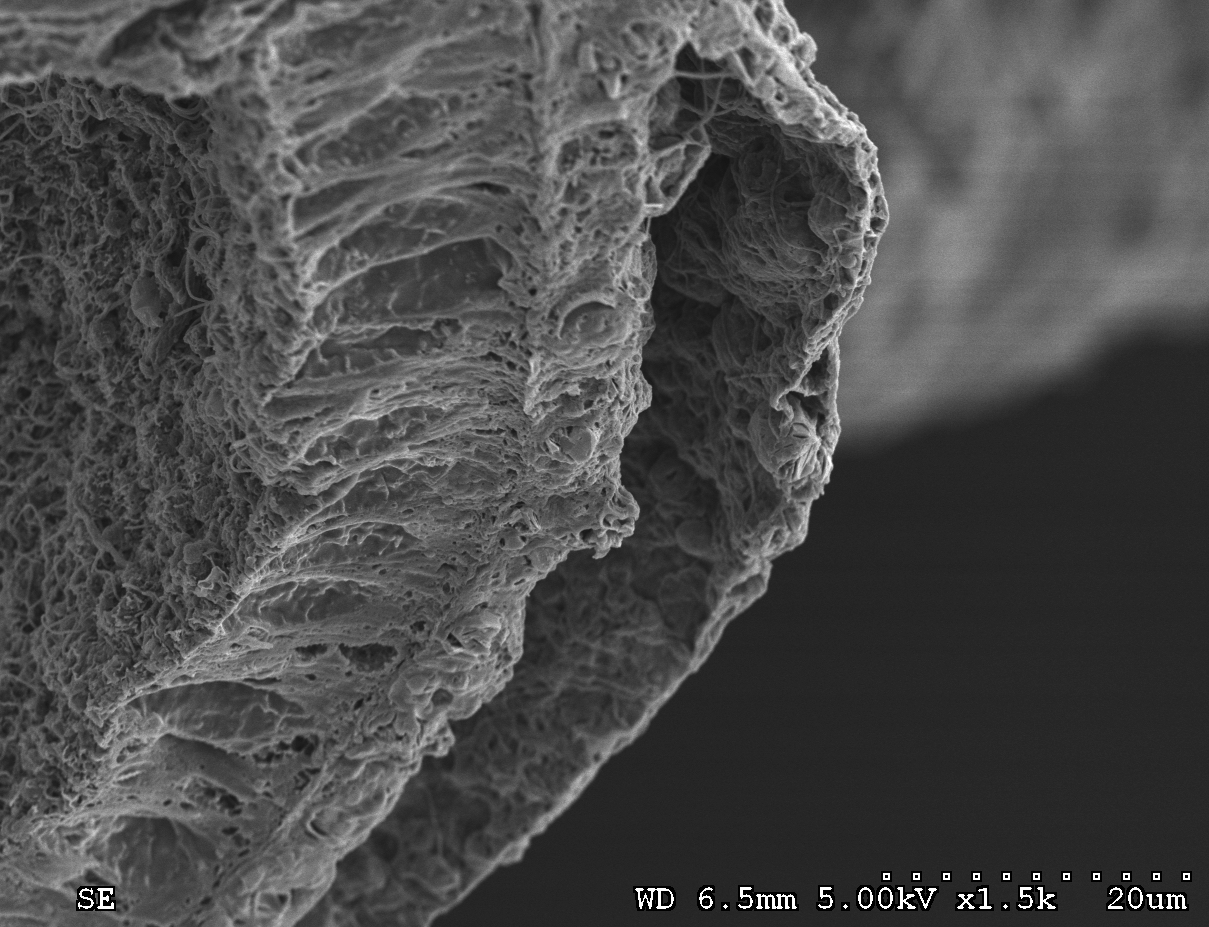
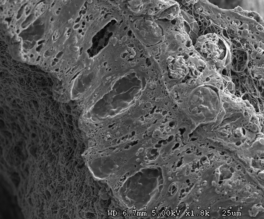
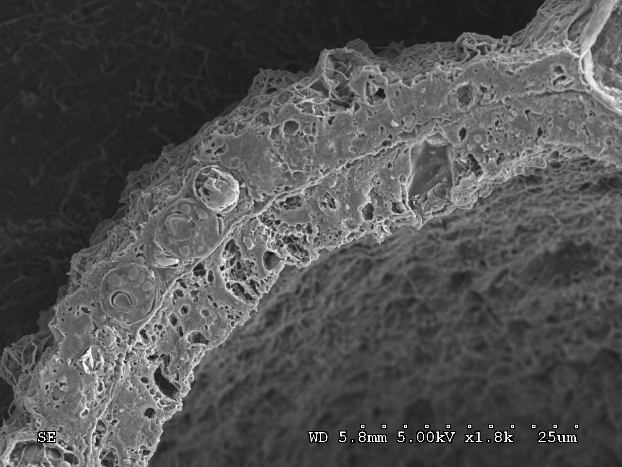
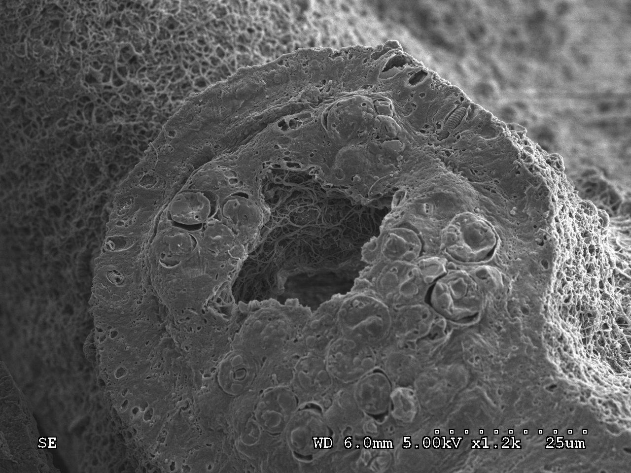
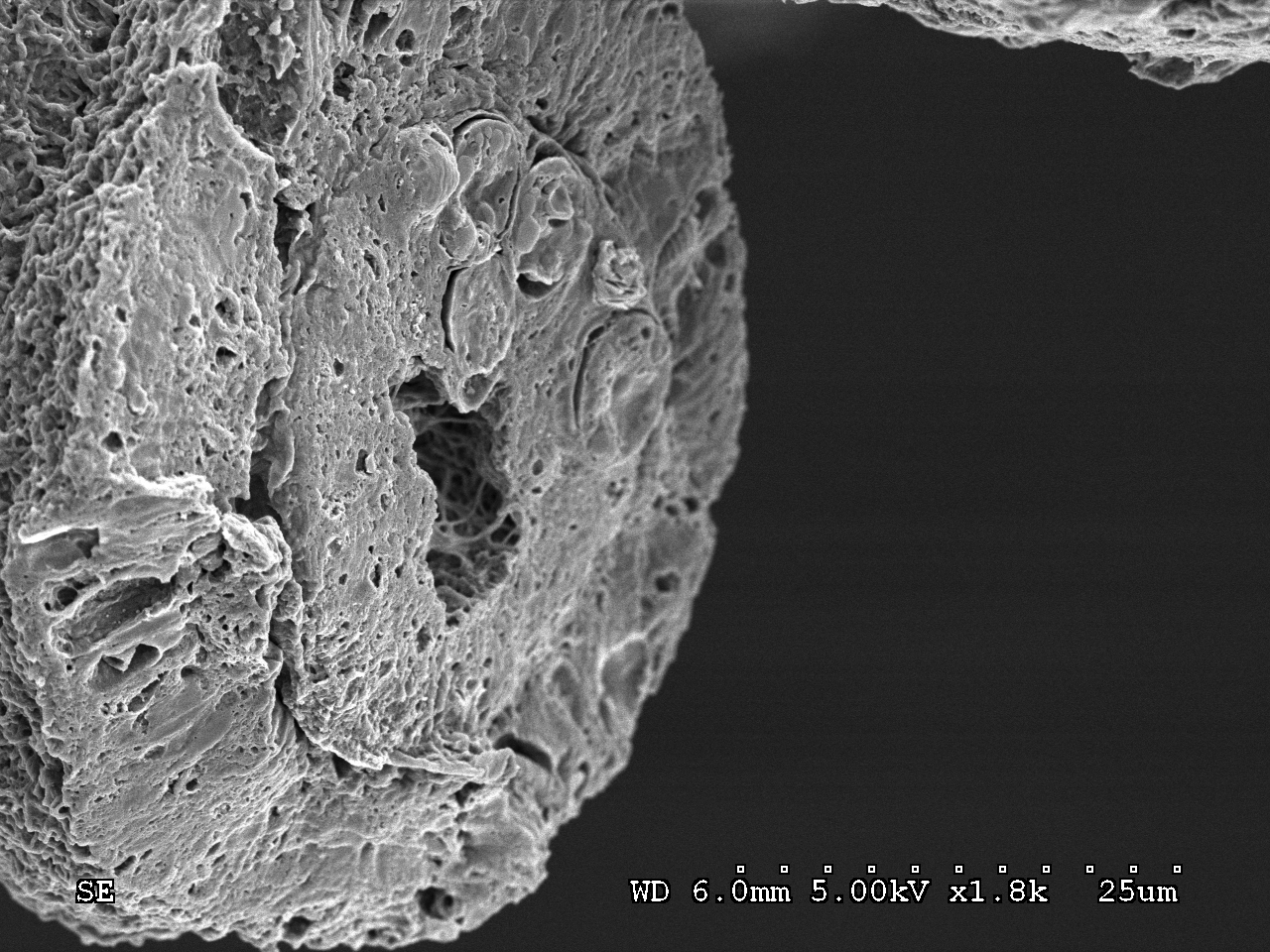
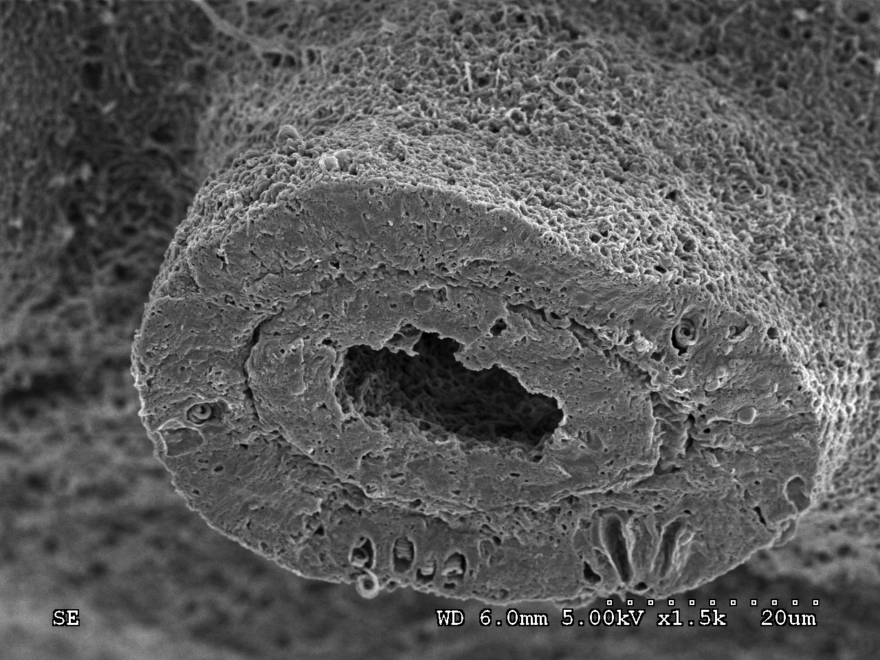
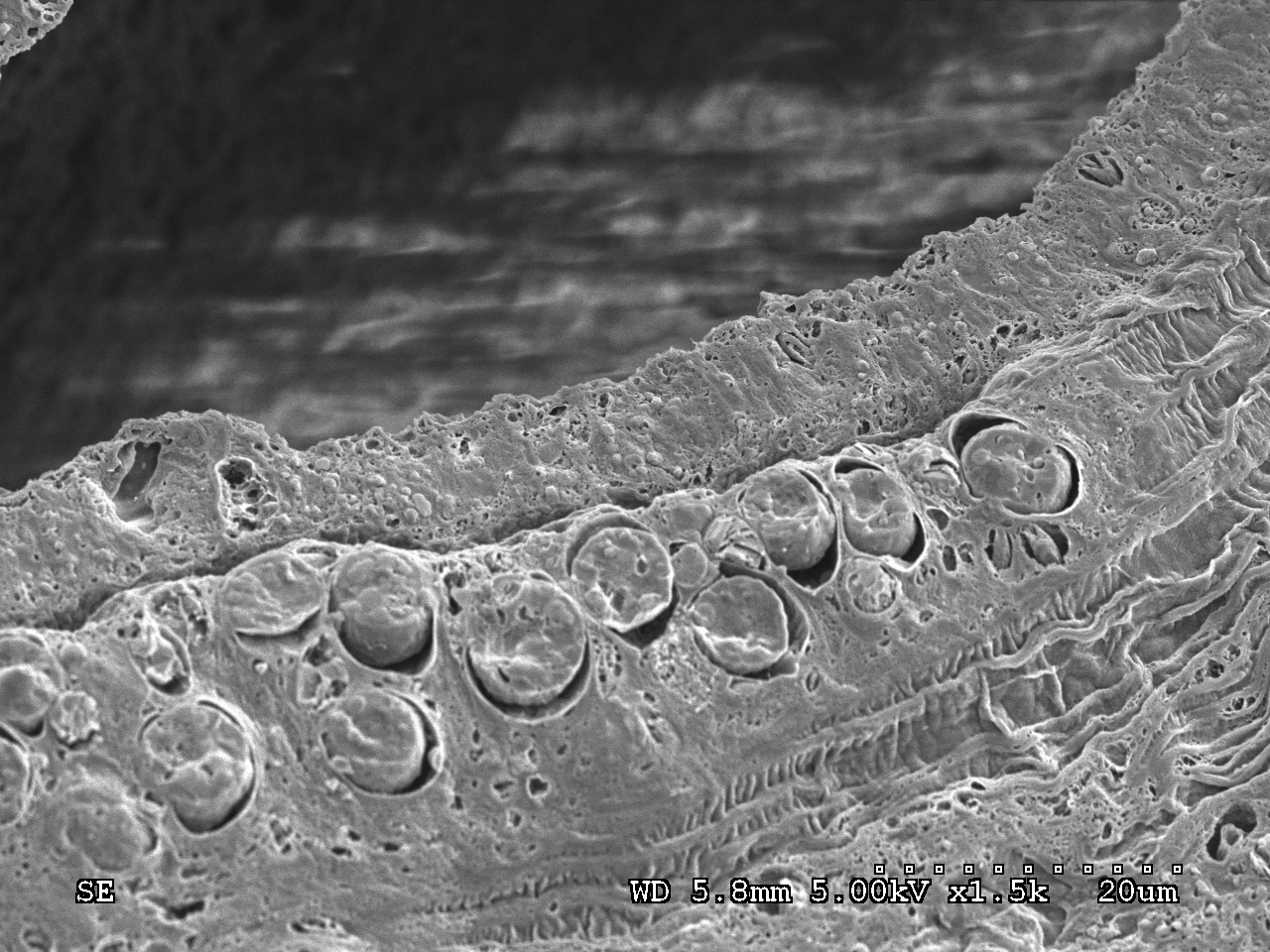
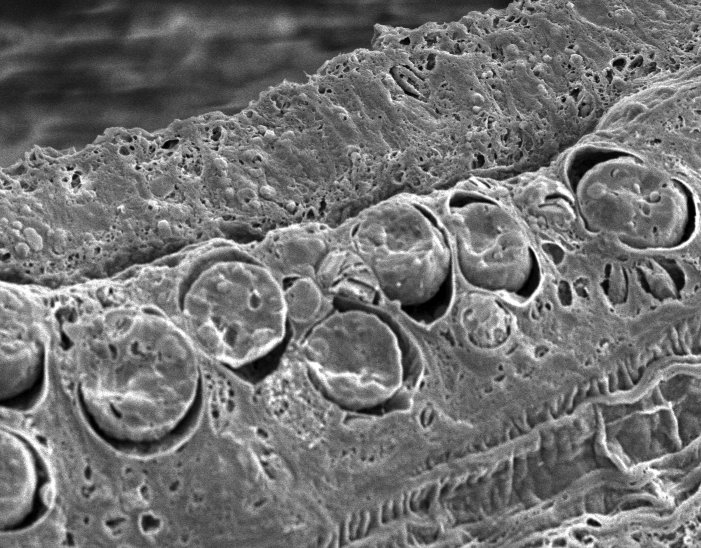
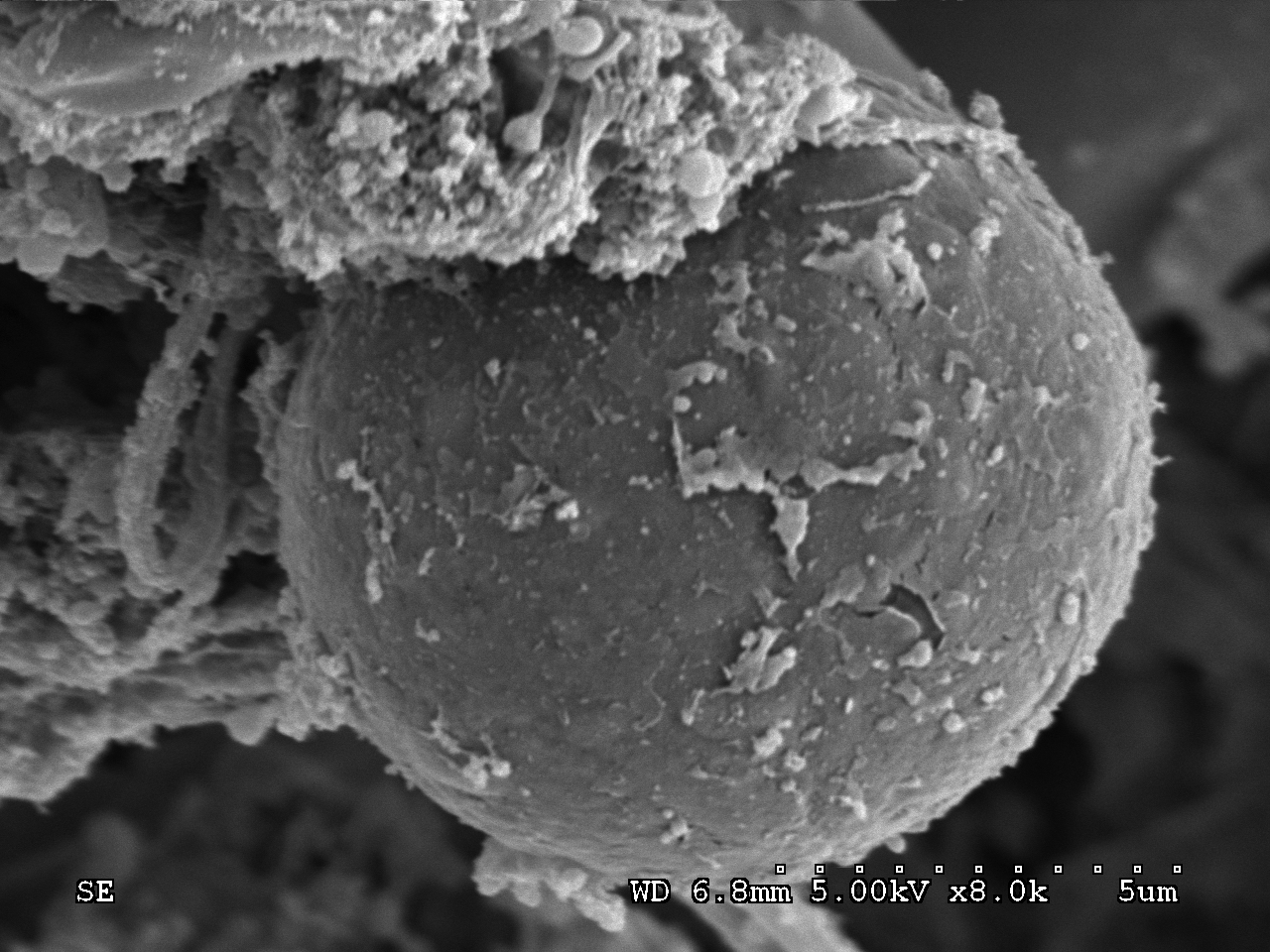
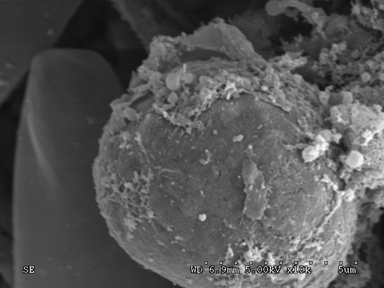
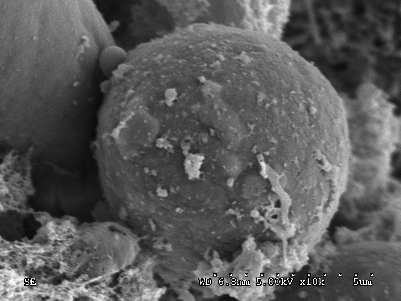
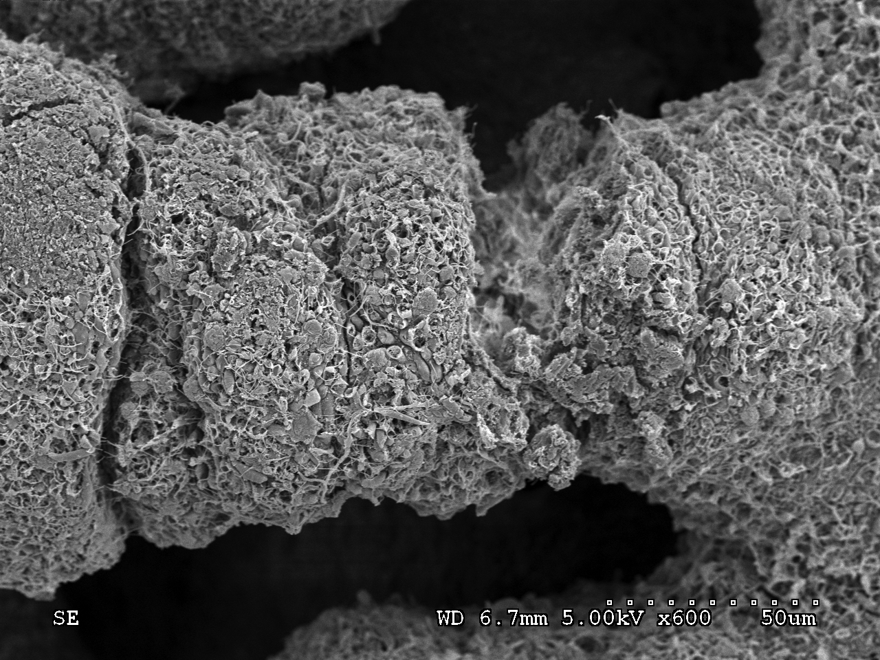
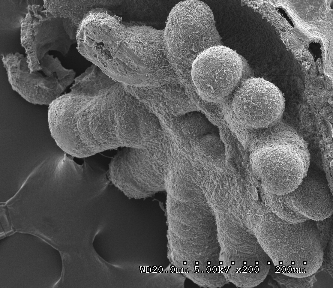
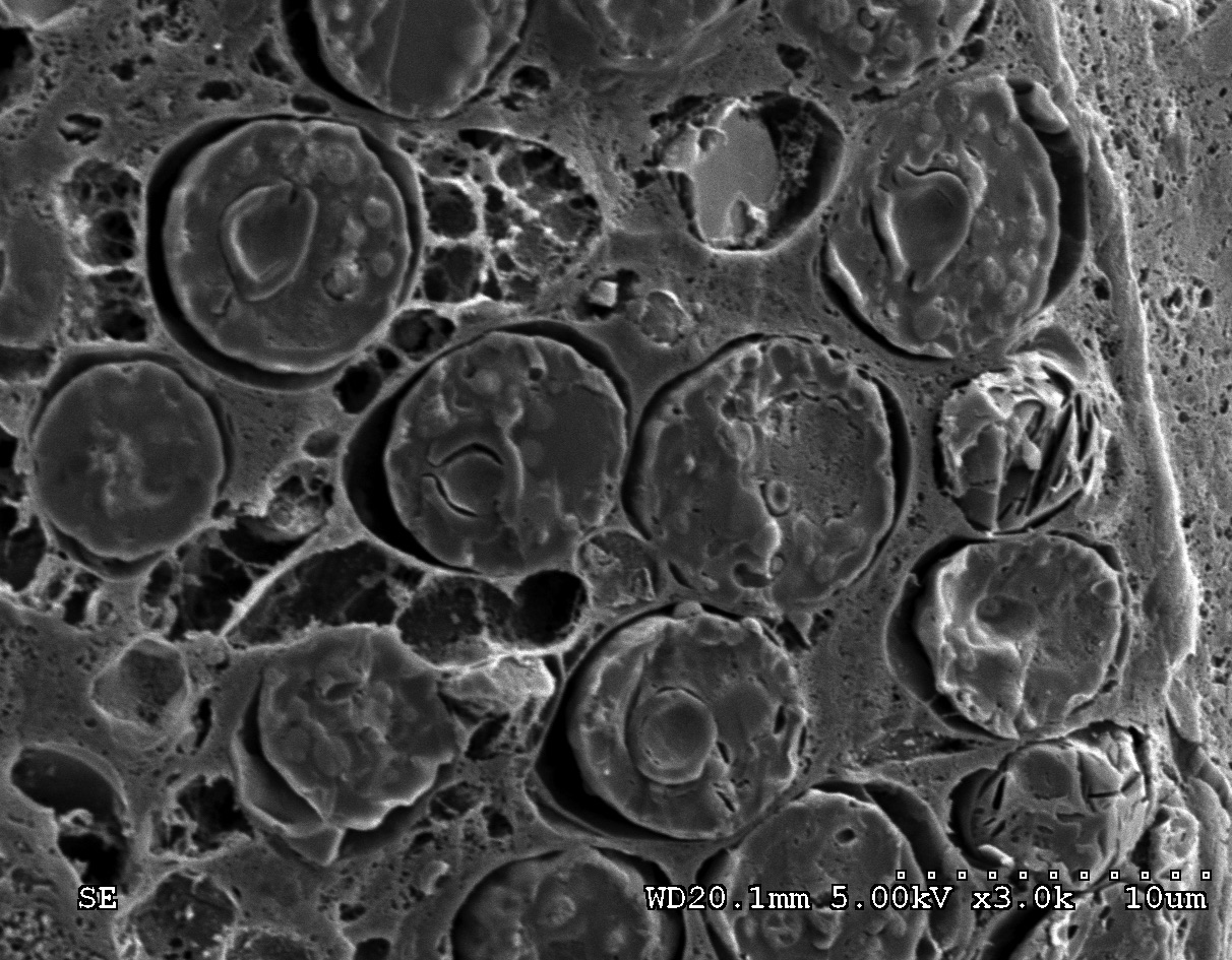
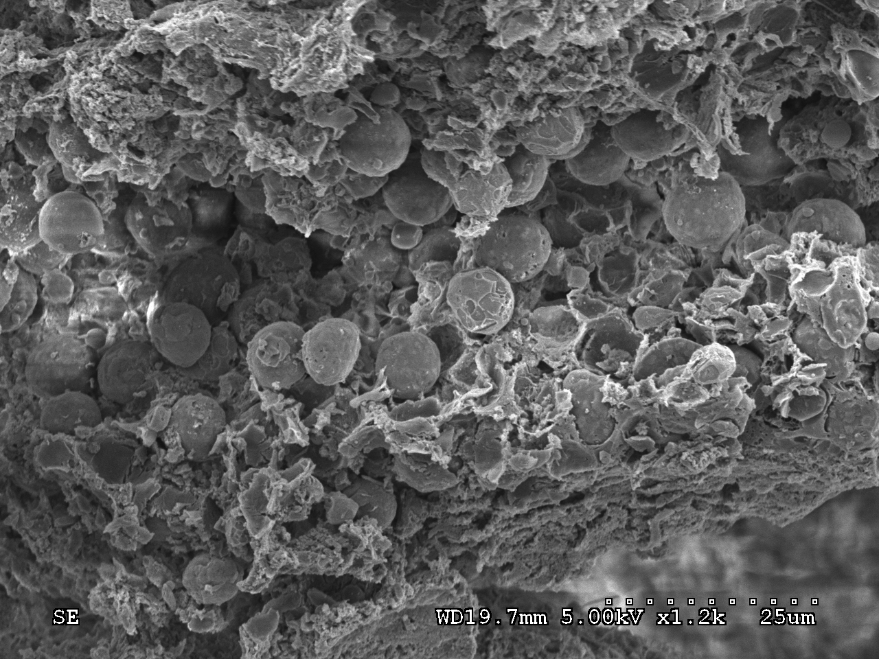
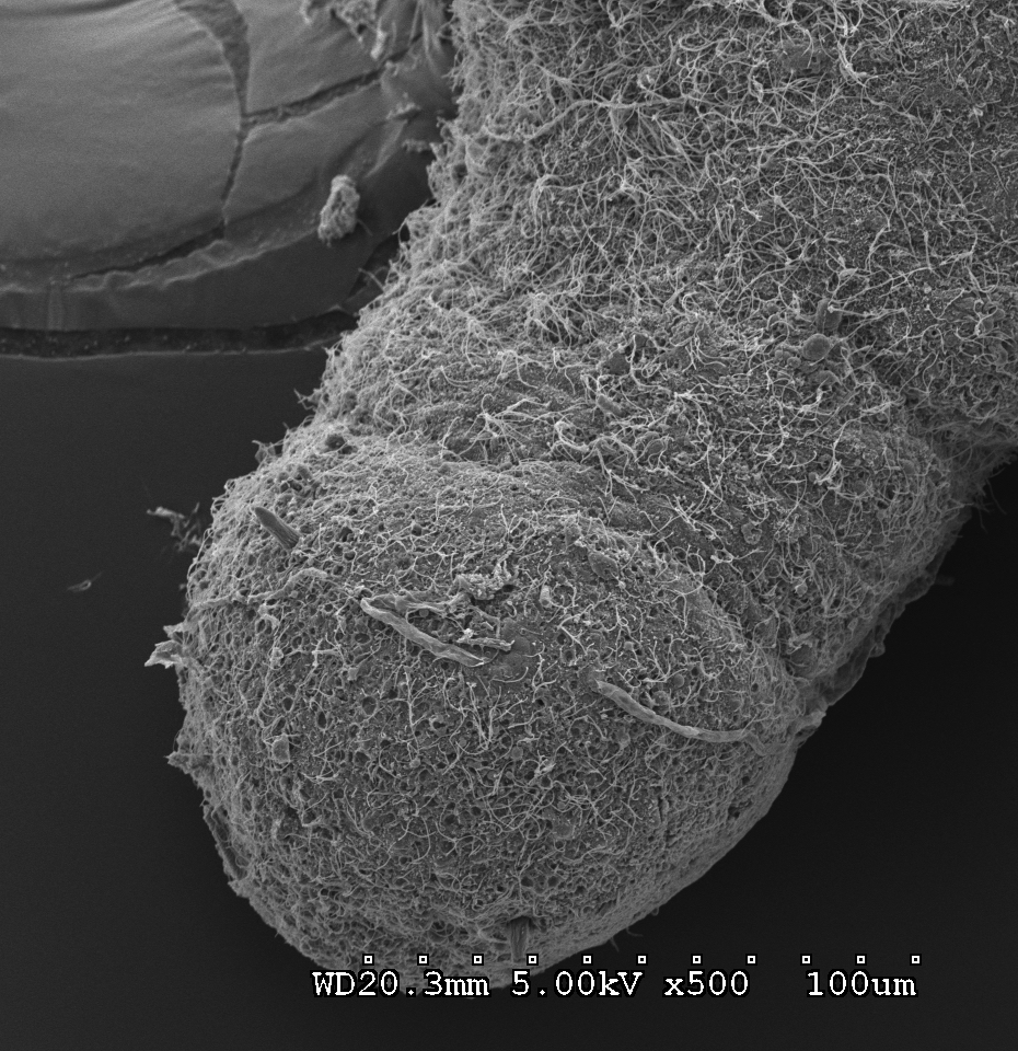
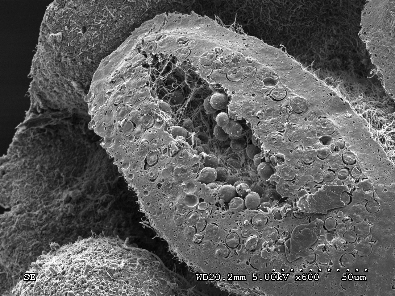
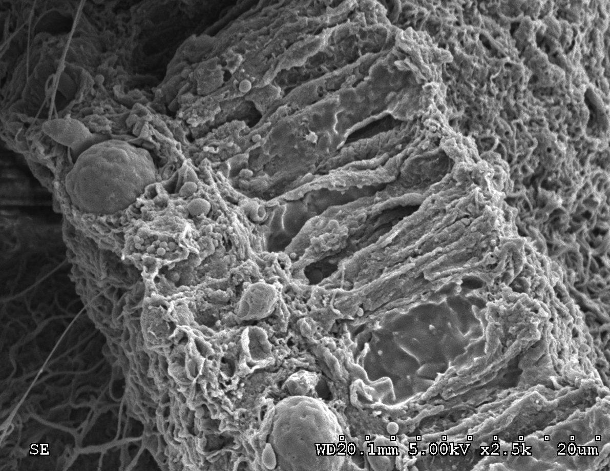
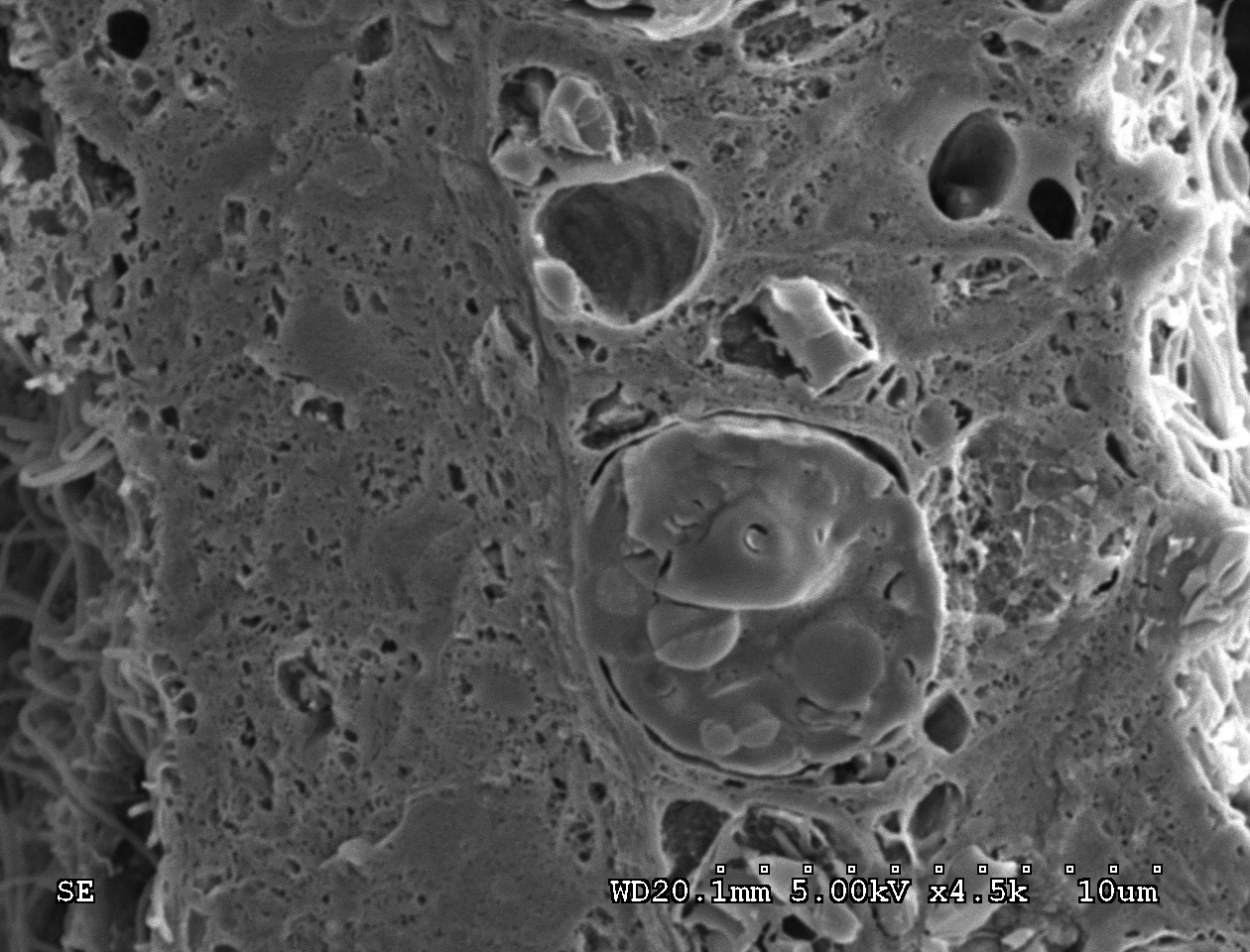
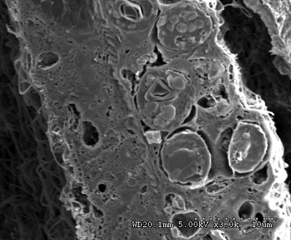
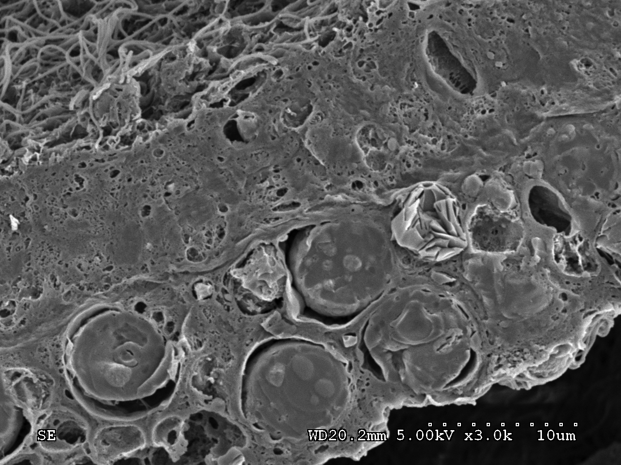
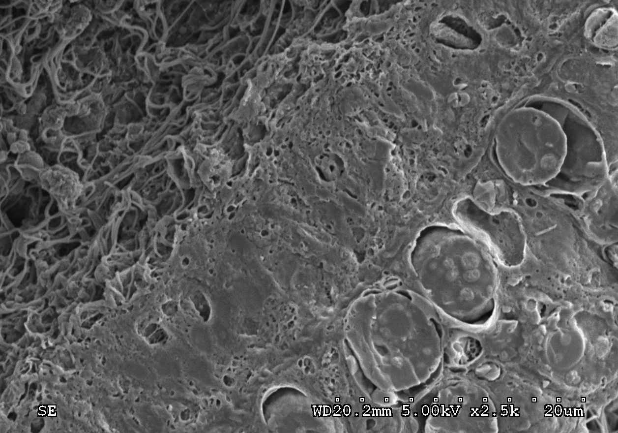
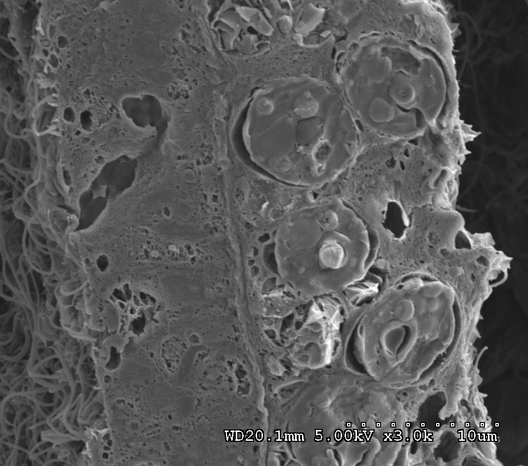
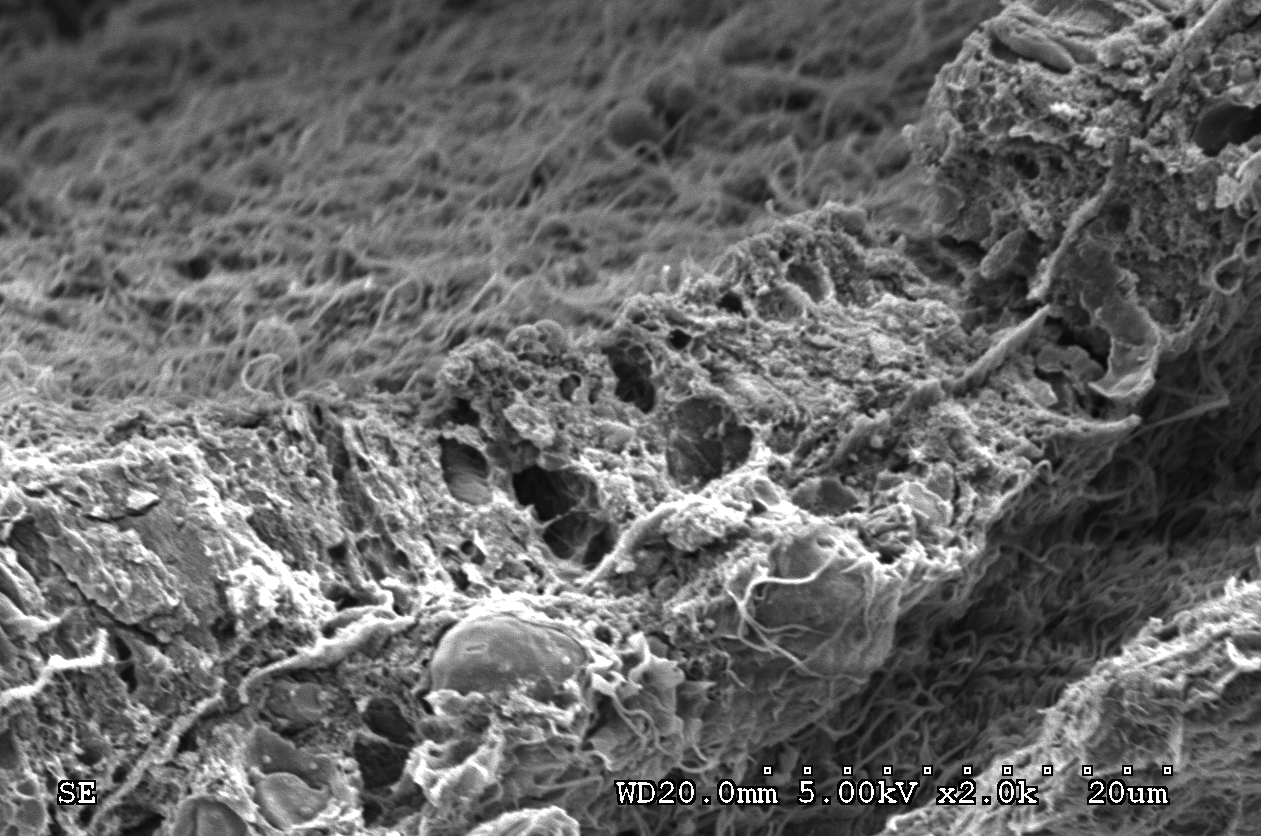
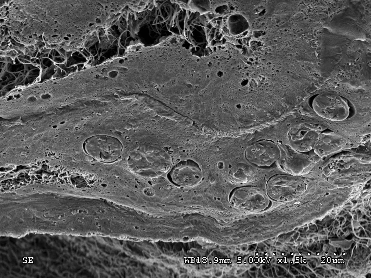
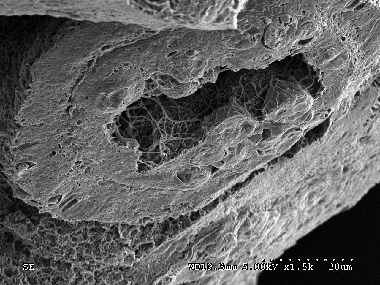
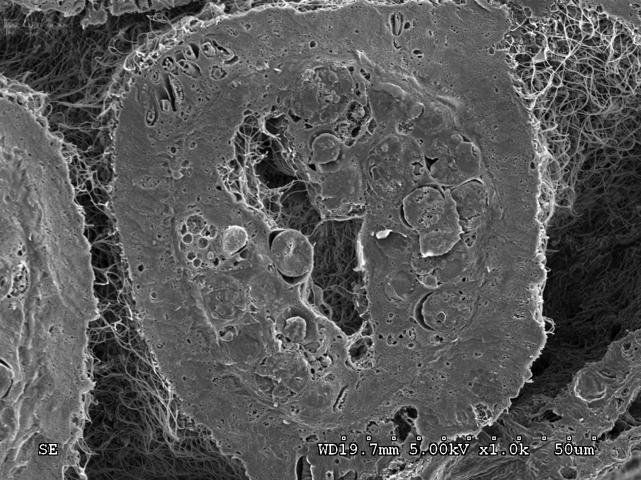
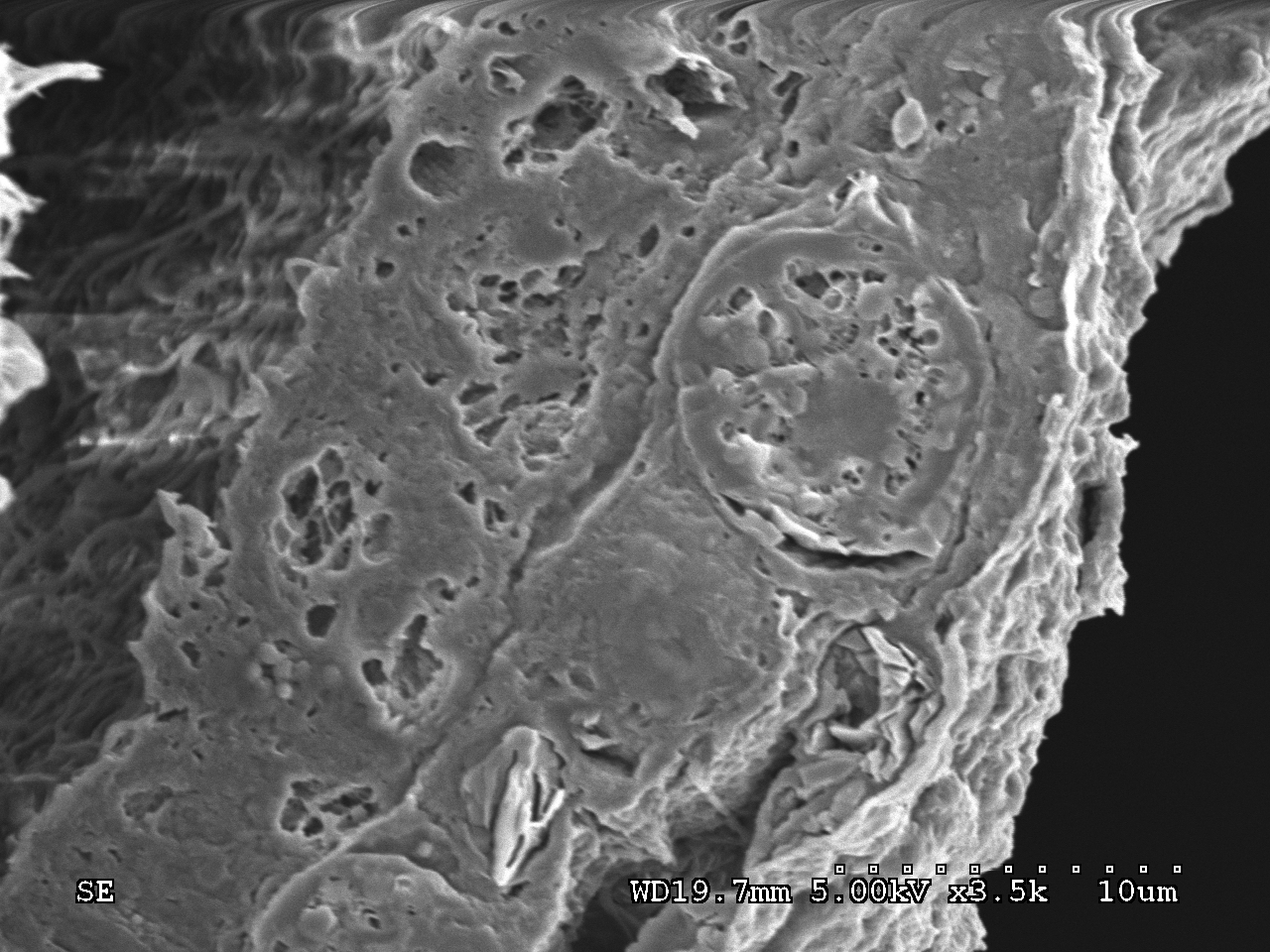
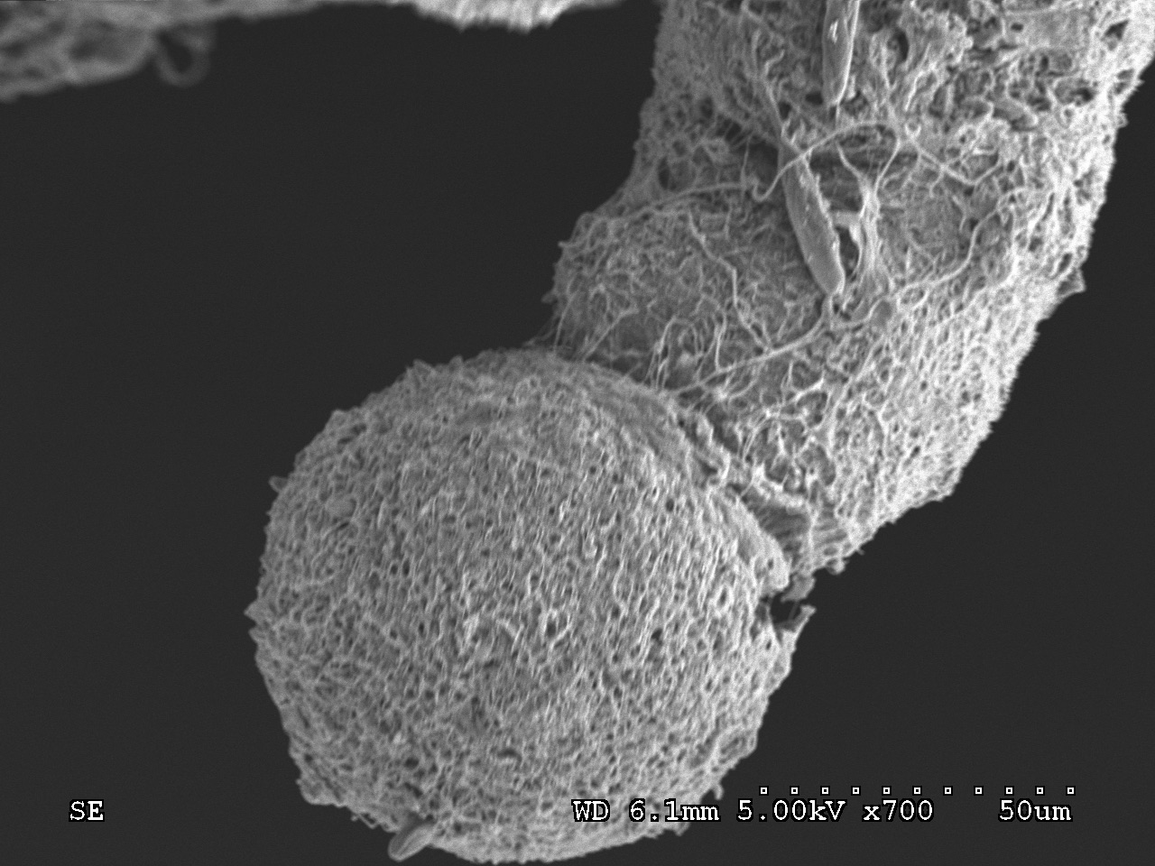
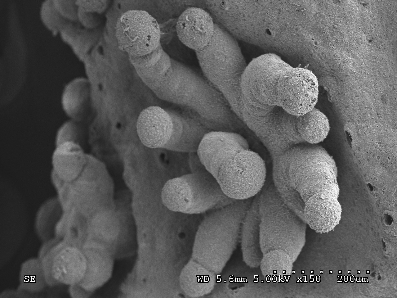
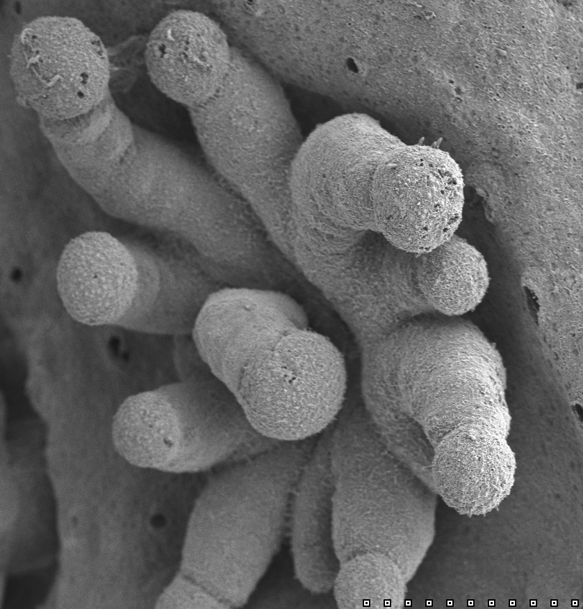
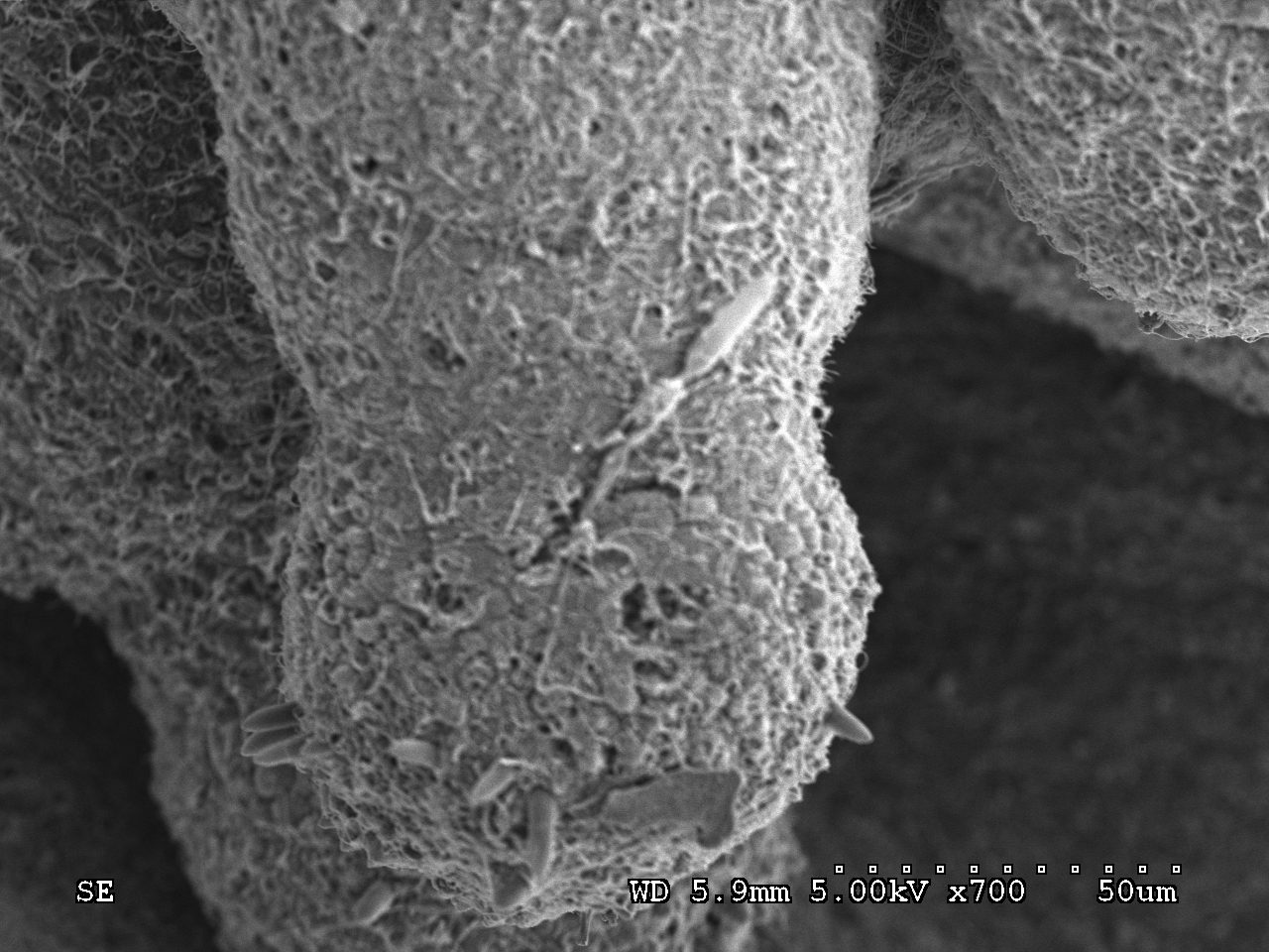
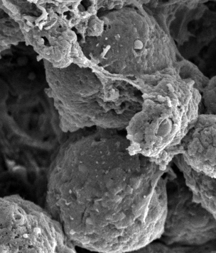
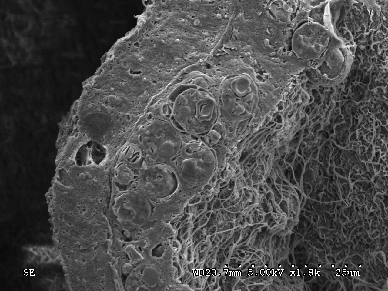
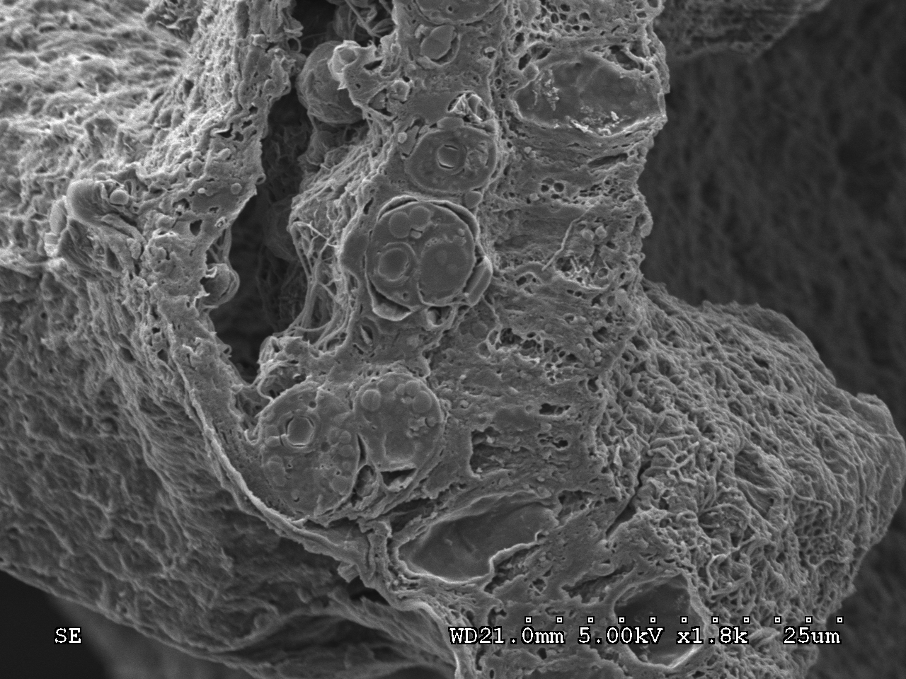
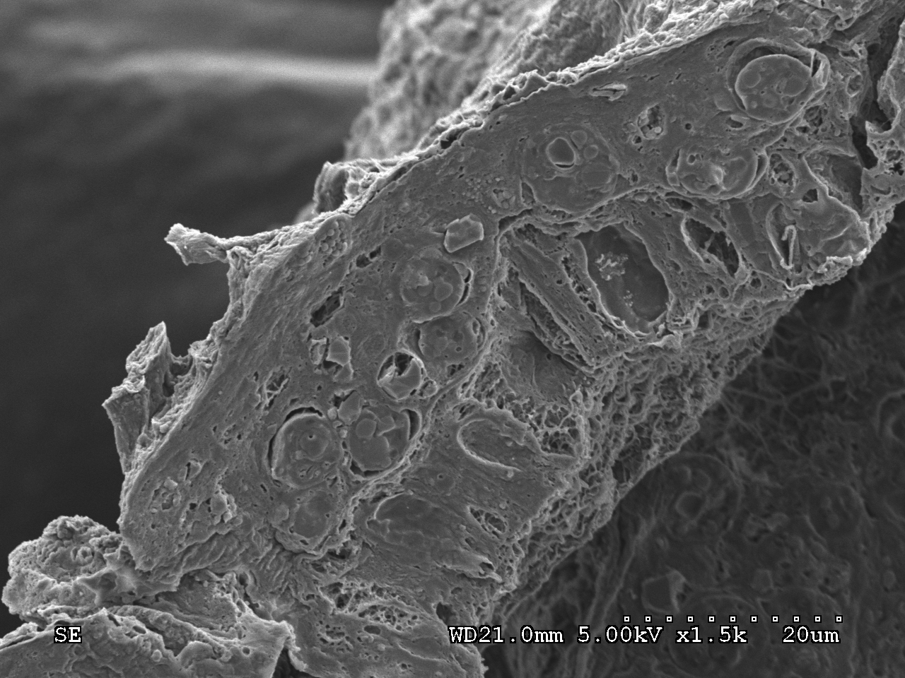
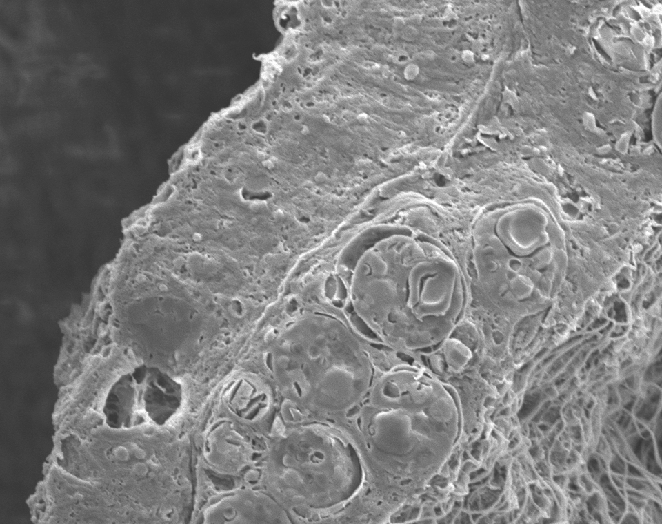
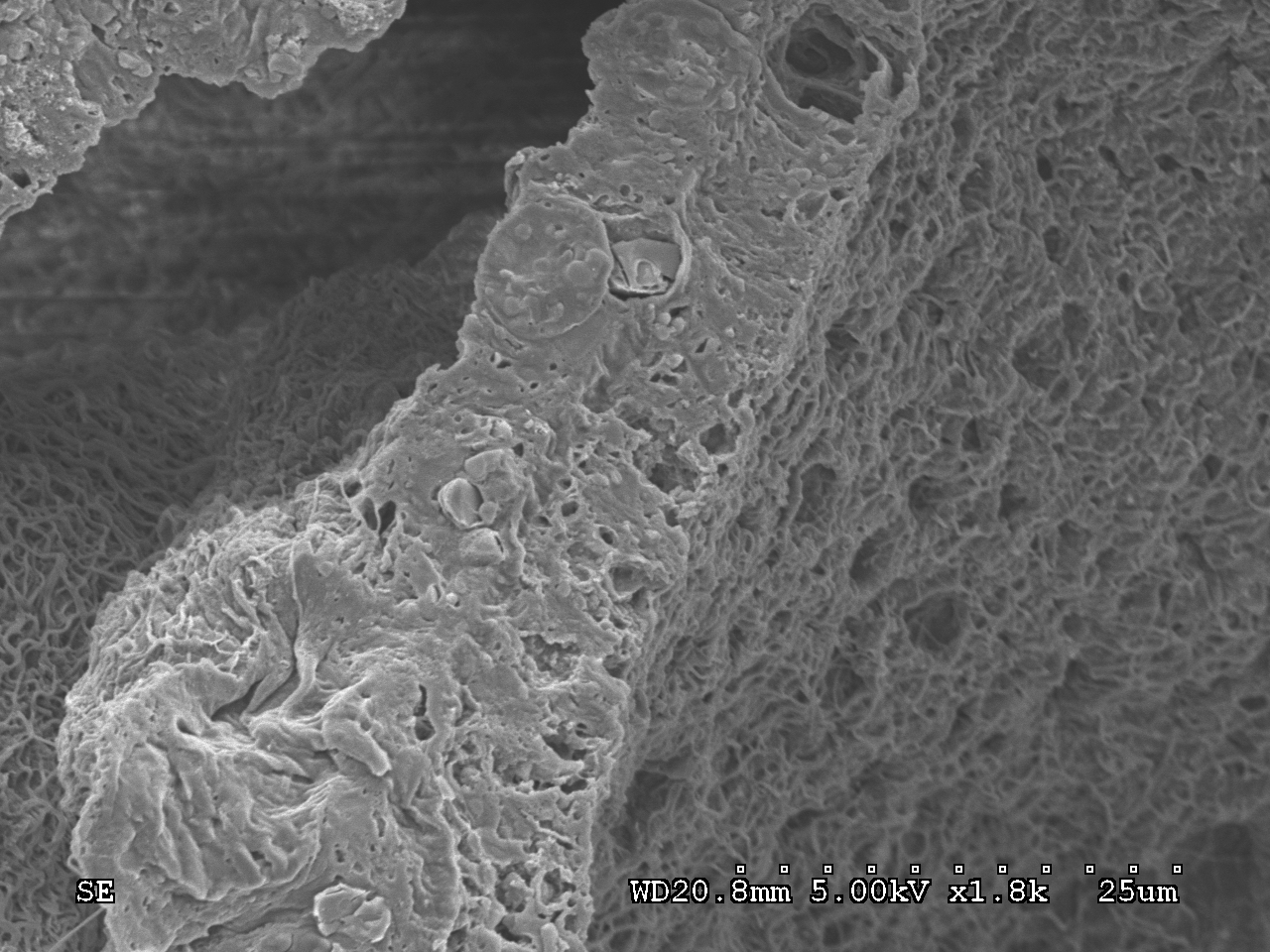
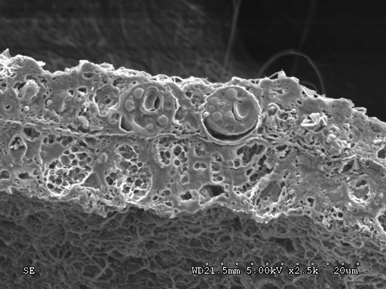
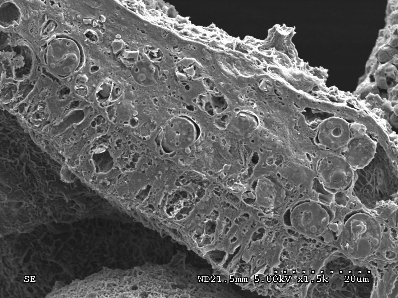
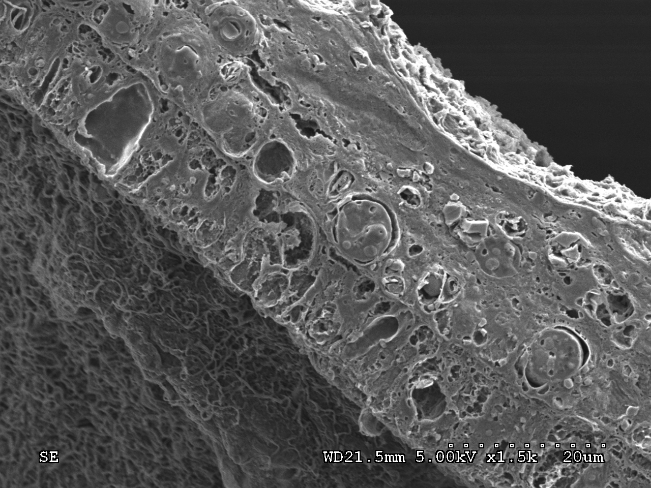
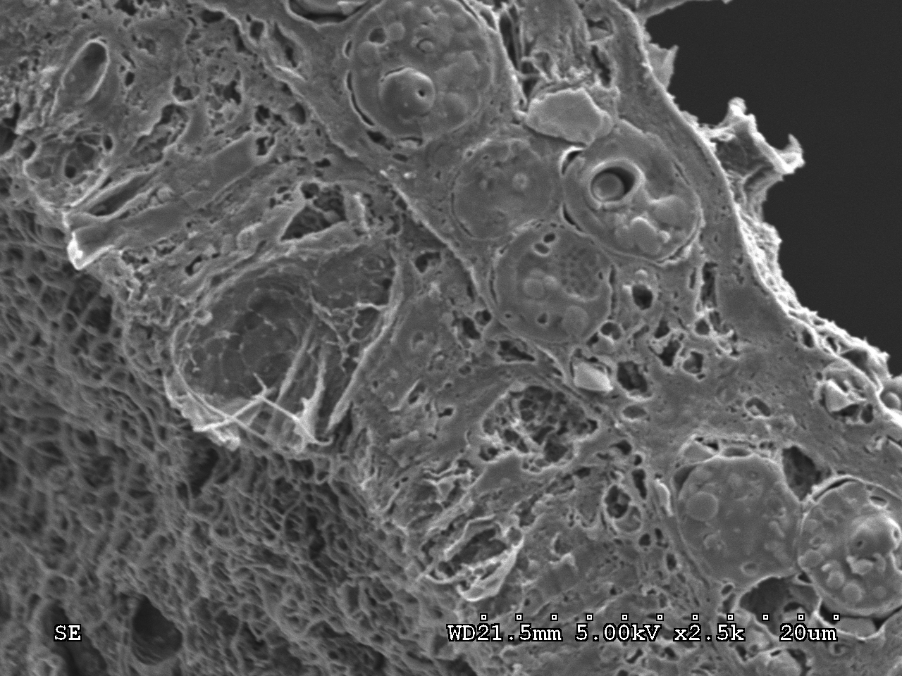
For details on preparation of Pocillopora damicornis samples for scanning electron microscopy (SEM), please see Mayfield et al. (2013, Coral Reefs).
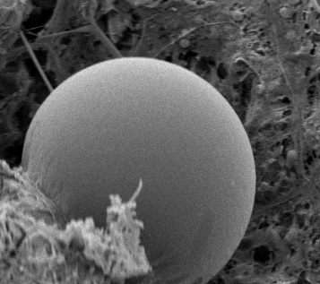
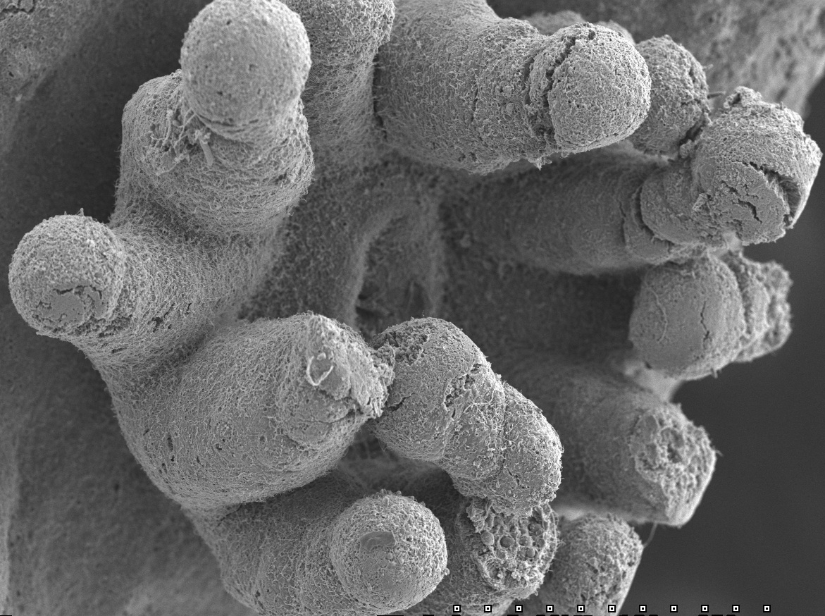
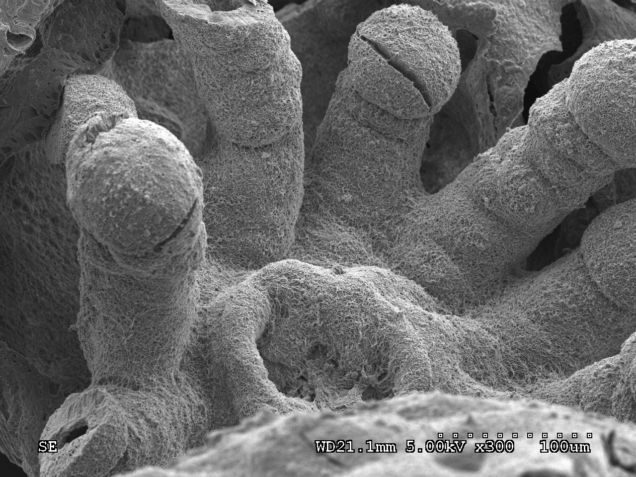
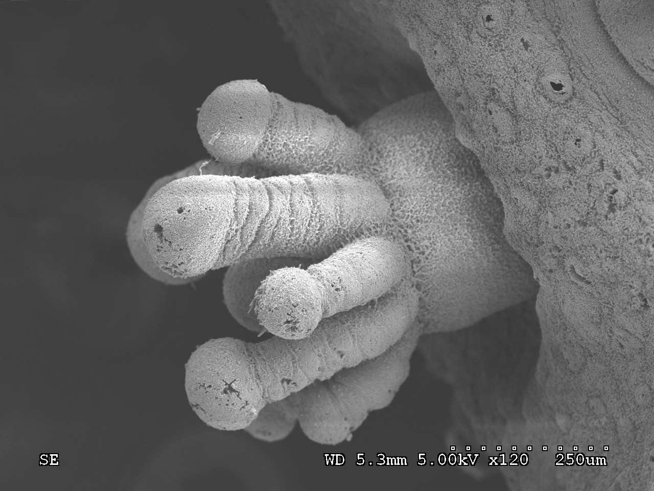
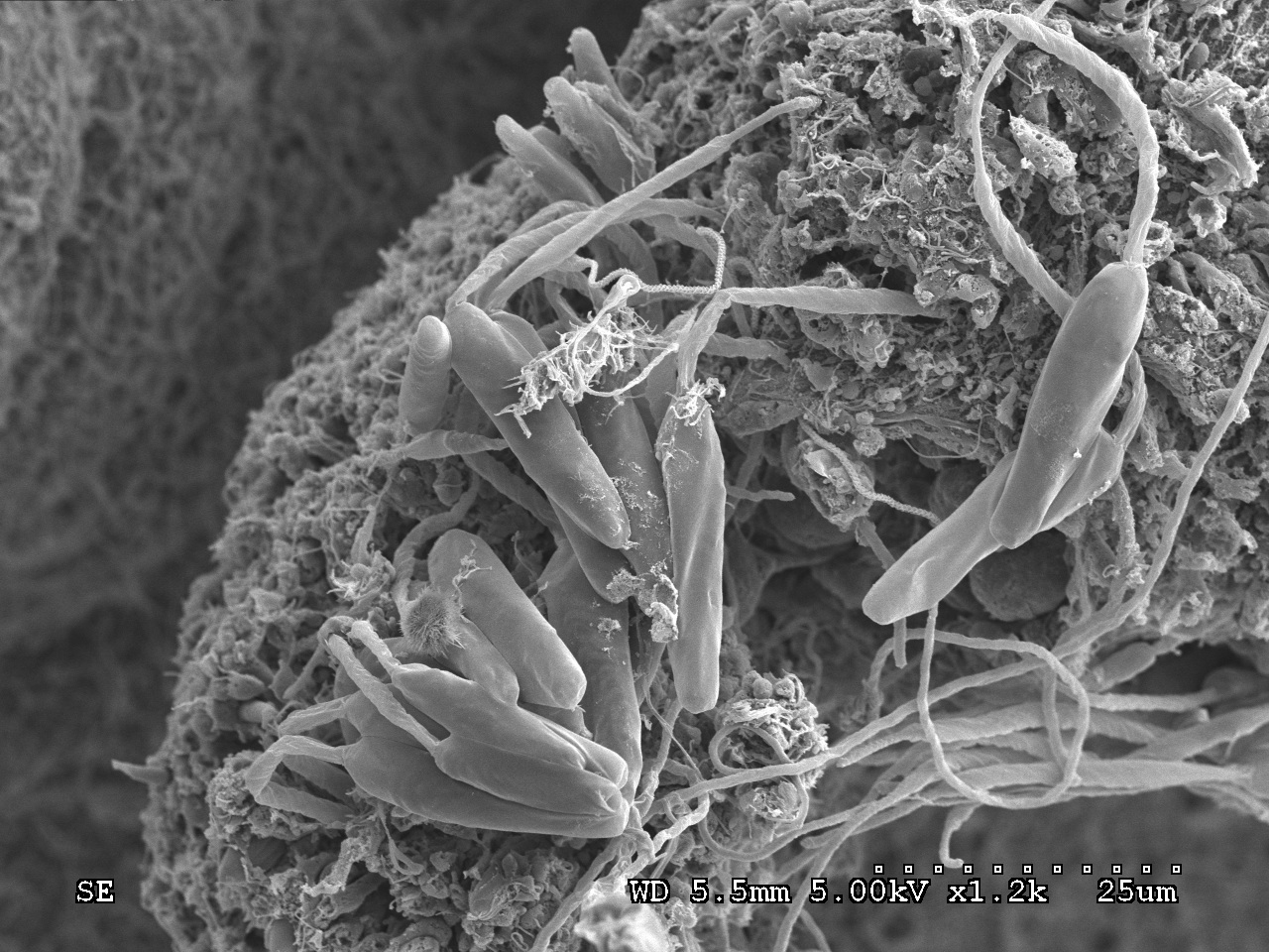
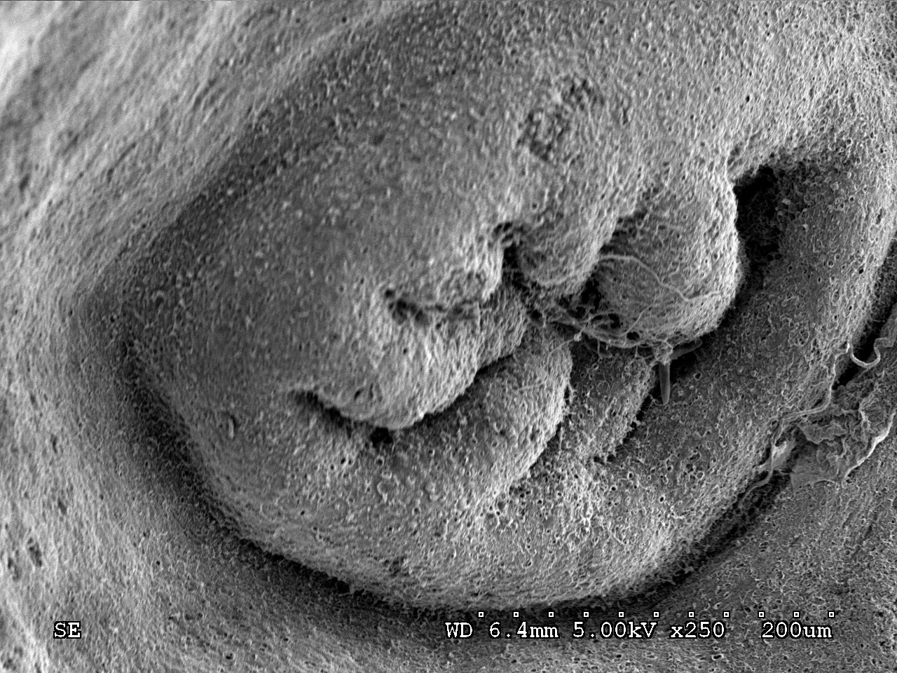
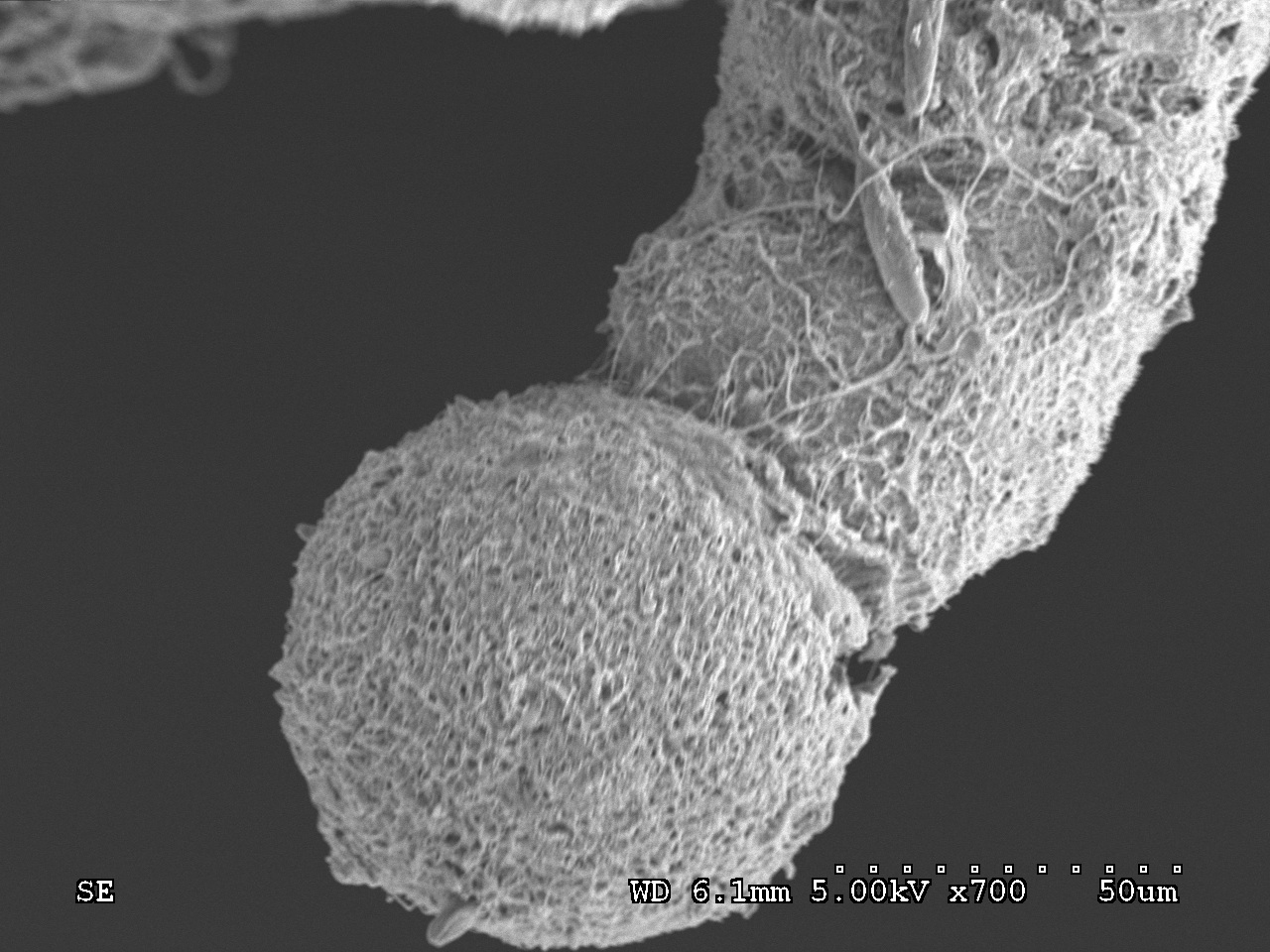
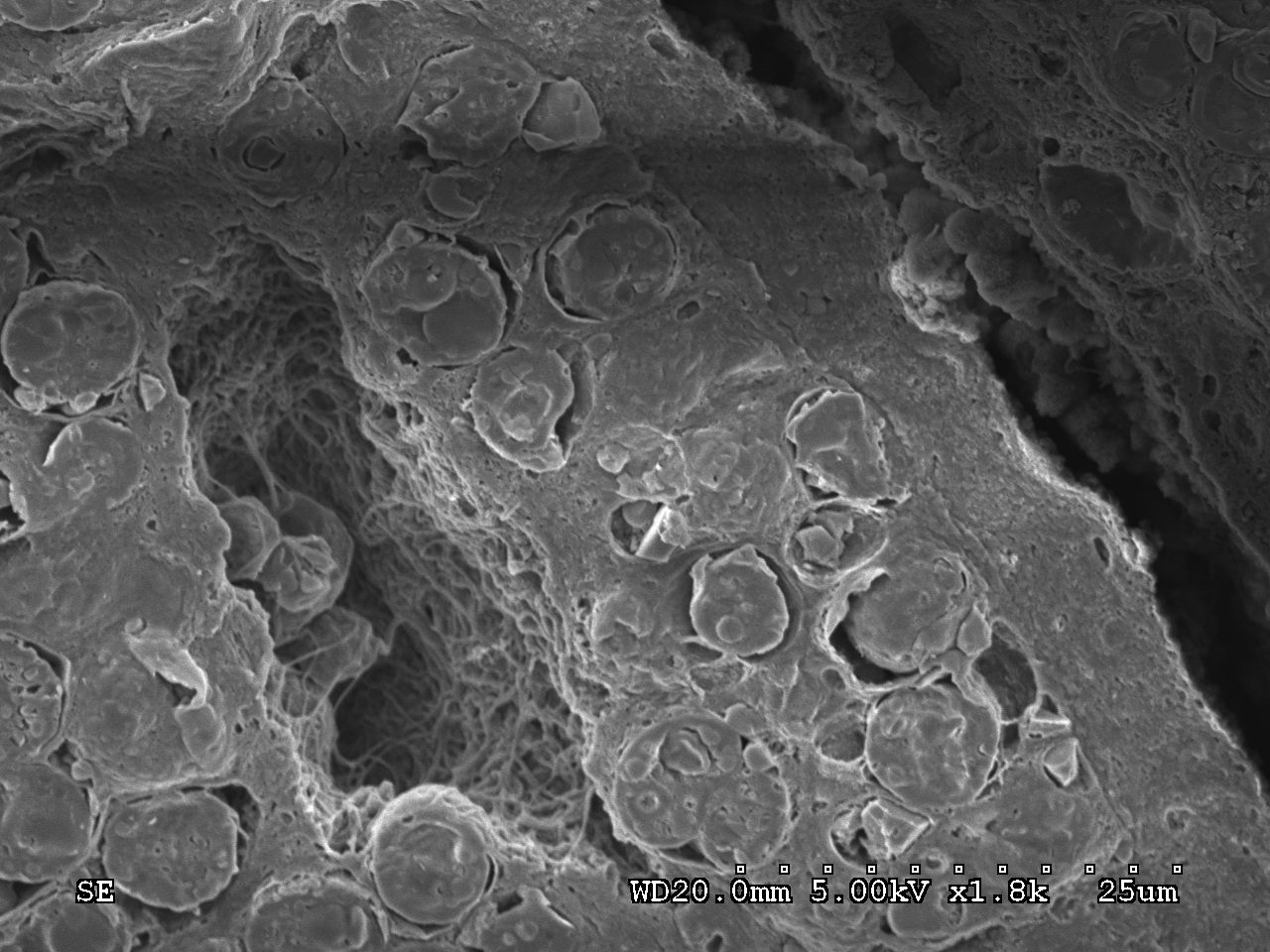
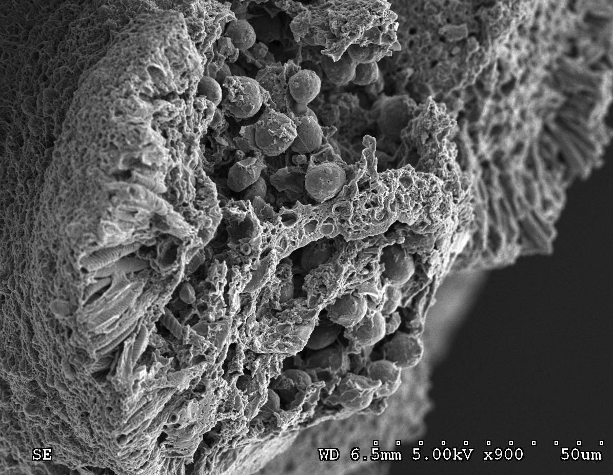
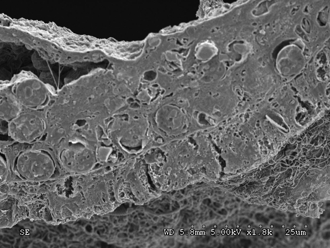
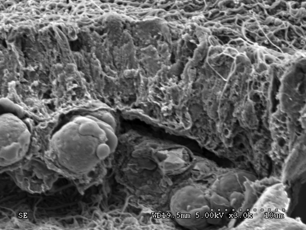
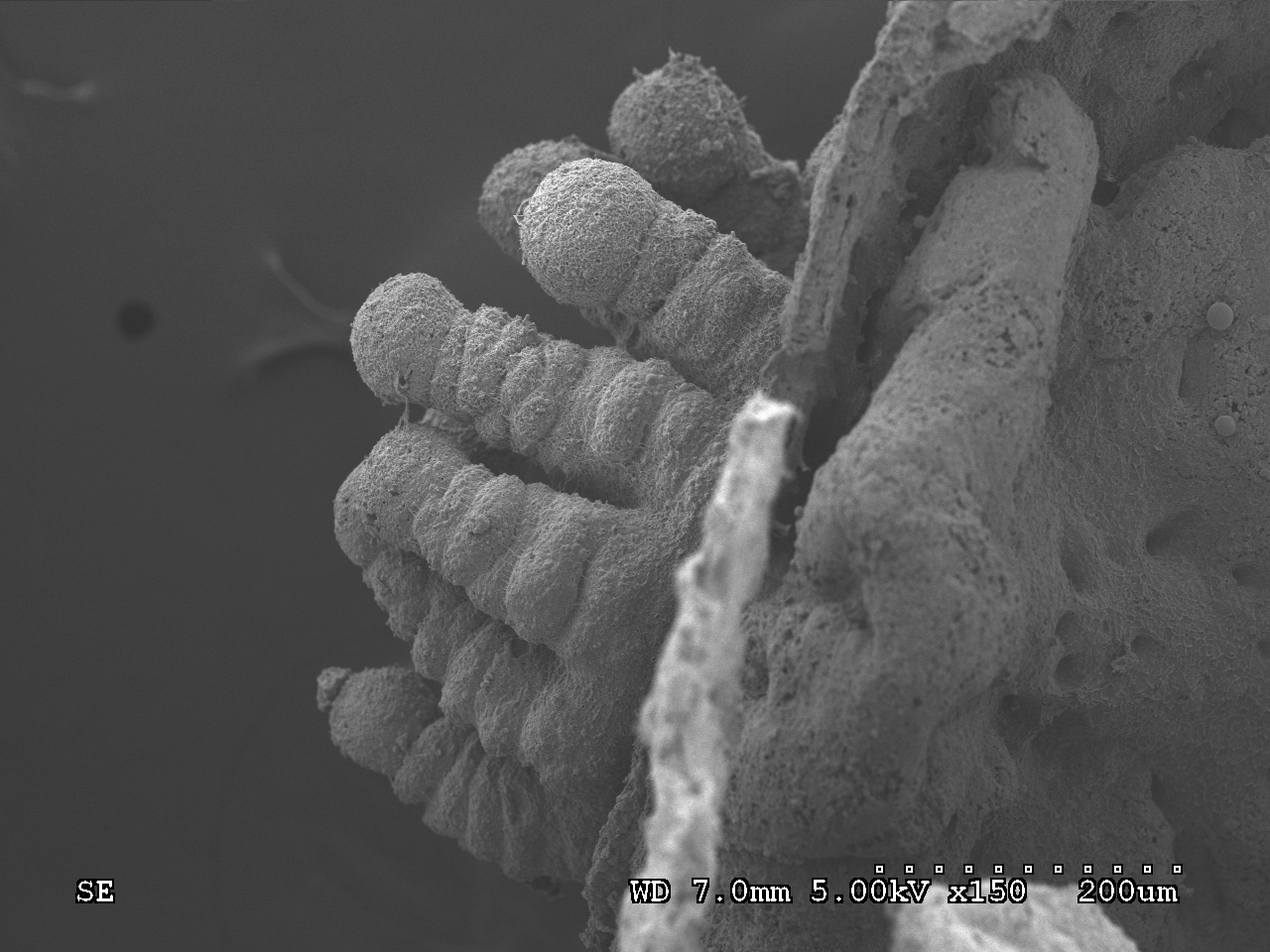
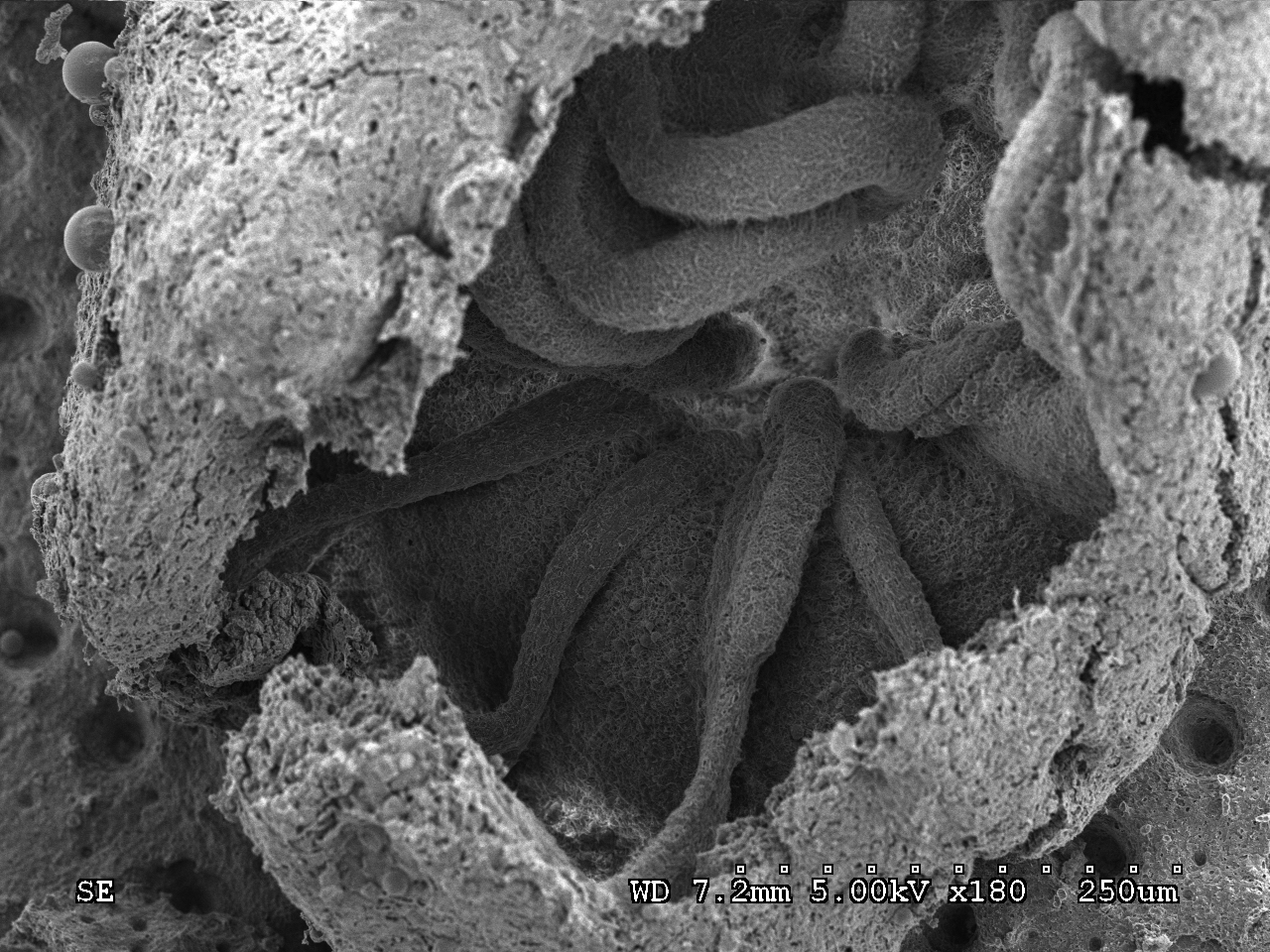
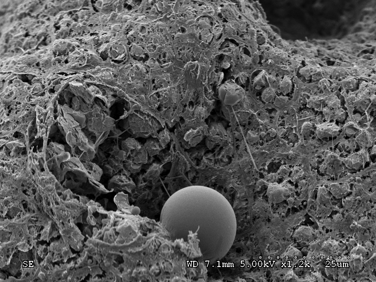
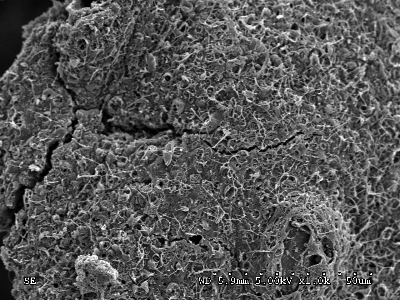
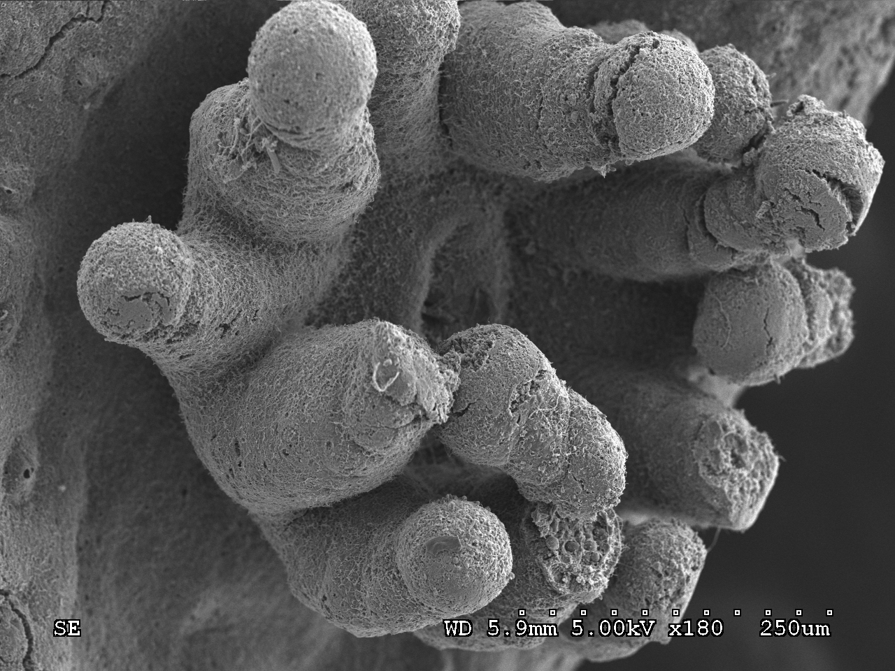
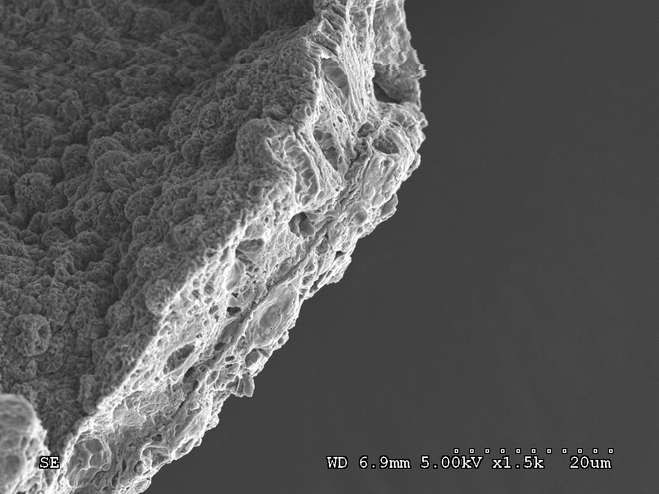
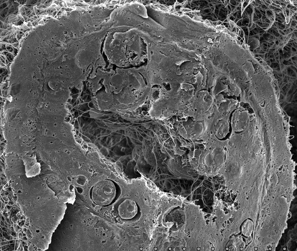
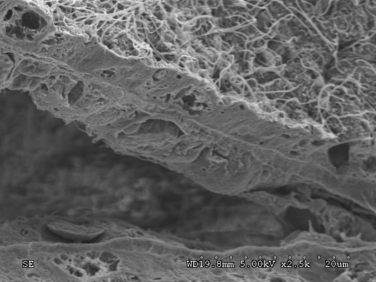
The images below represent tissues that were freeze fractured in liquid nitrogen. This allows for better visualization of the two tissue layers: the Symbiodinaceae-free epiderm and the gastroderm containing these endosymbiotic dinoflagellates.

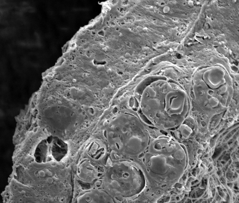
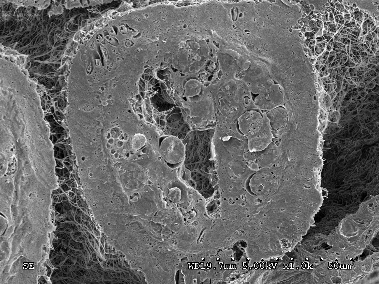
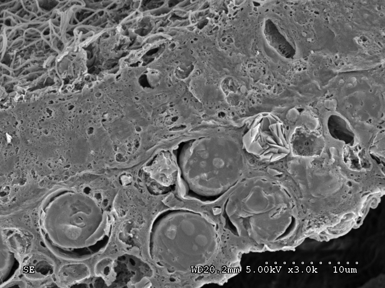
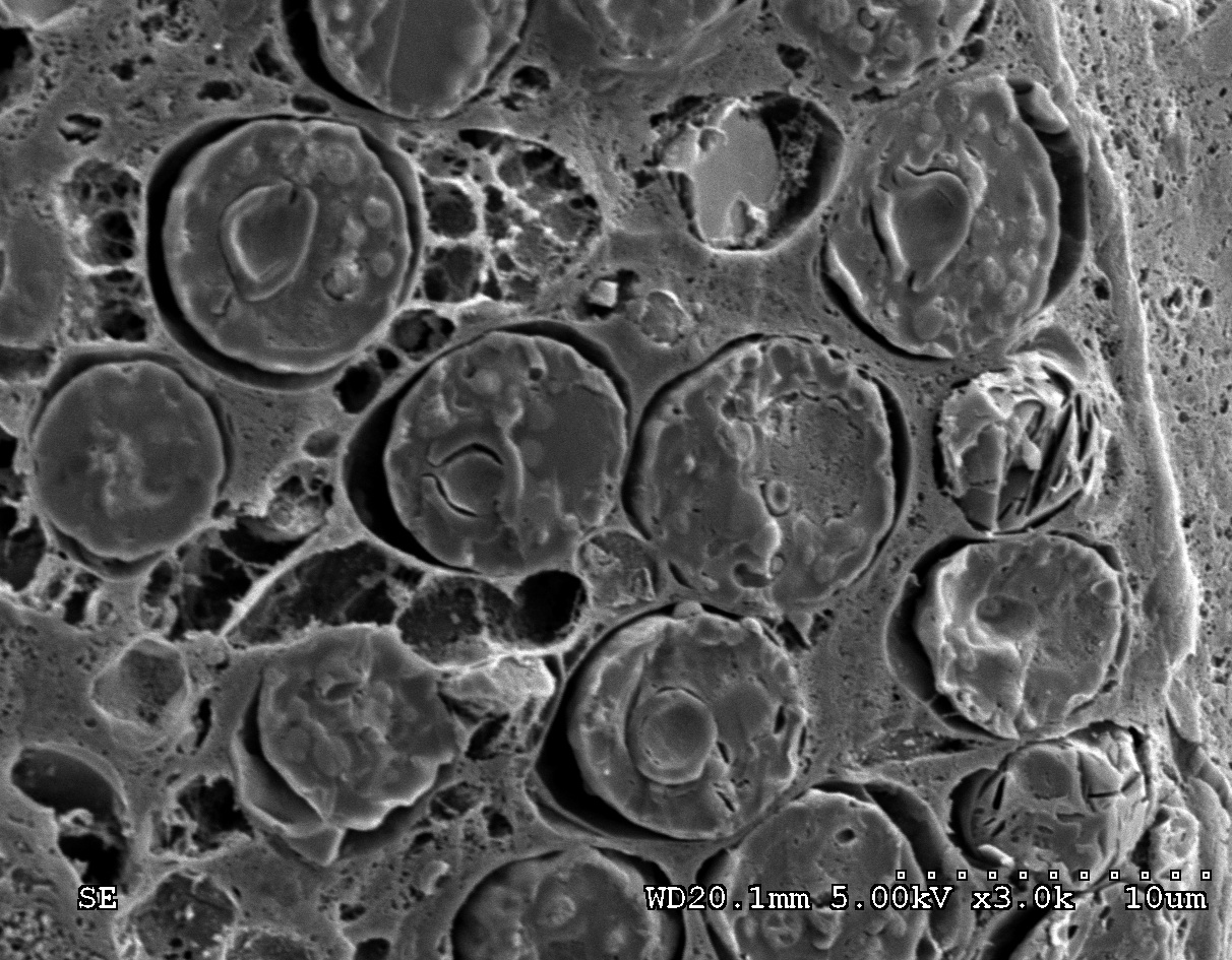
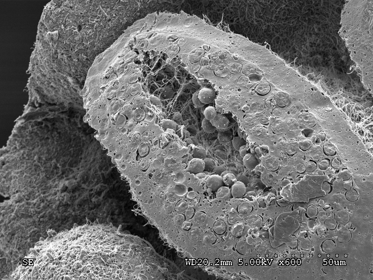
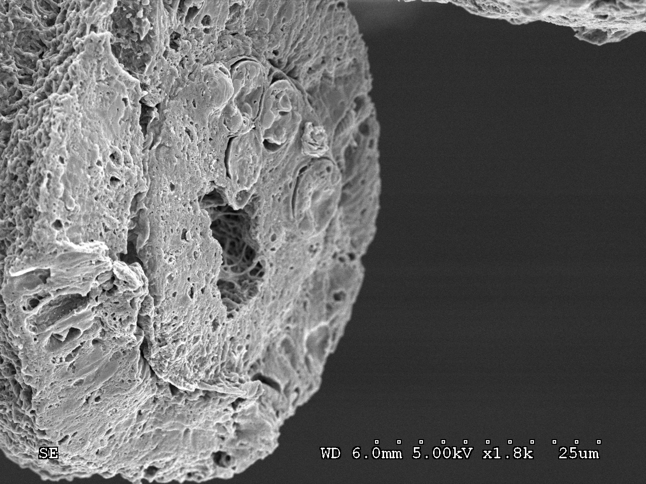
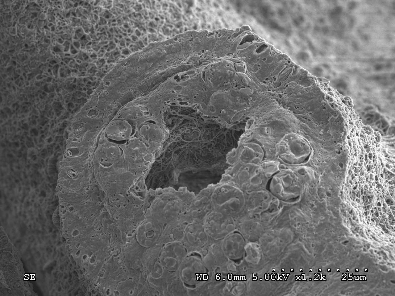
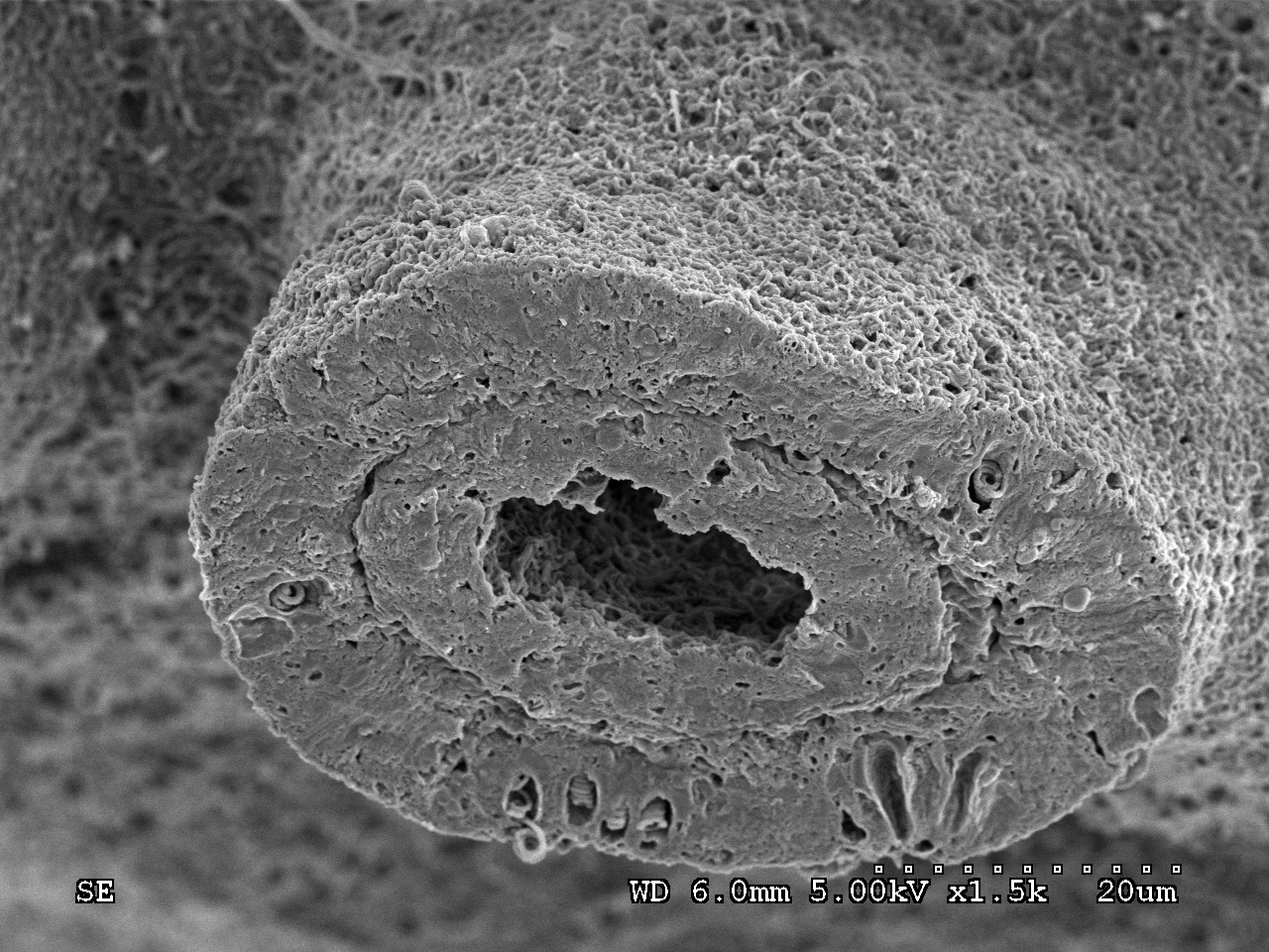
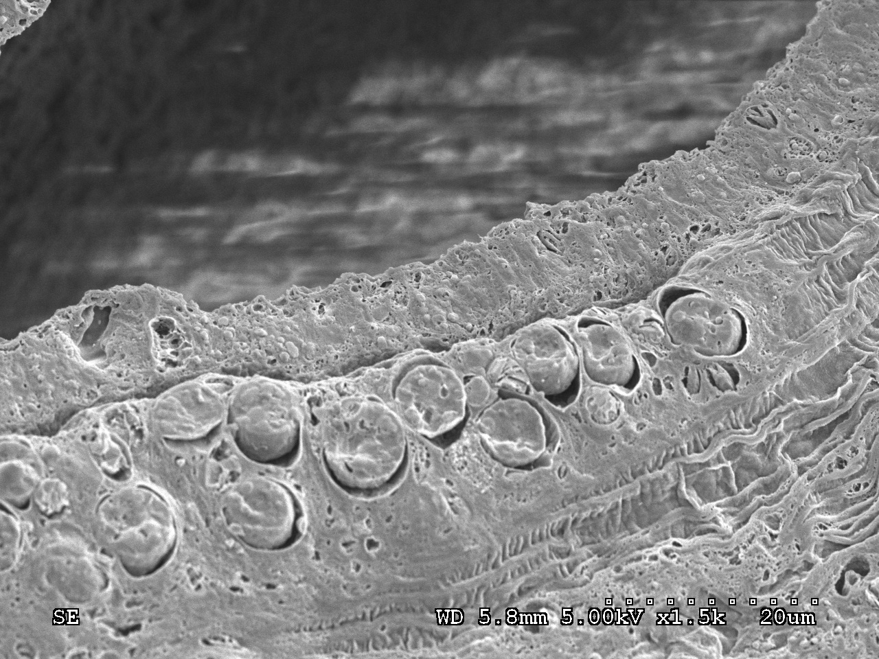
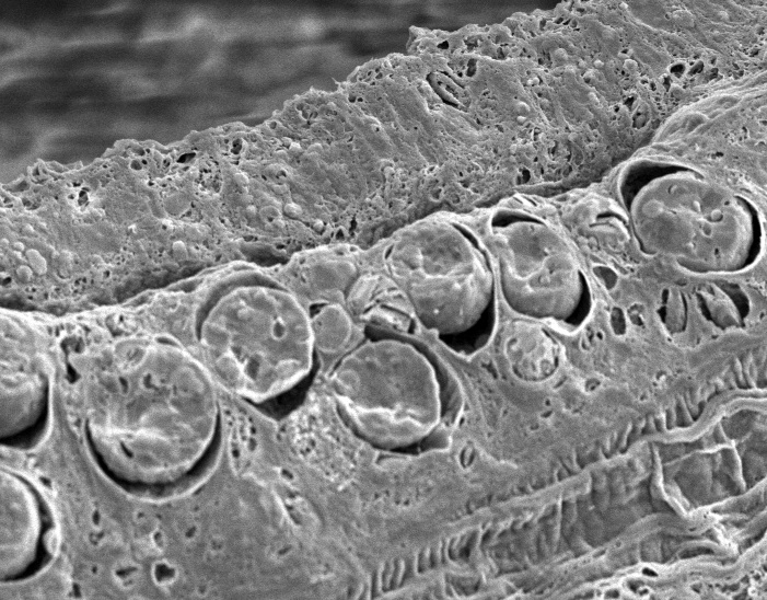
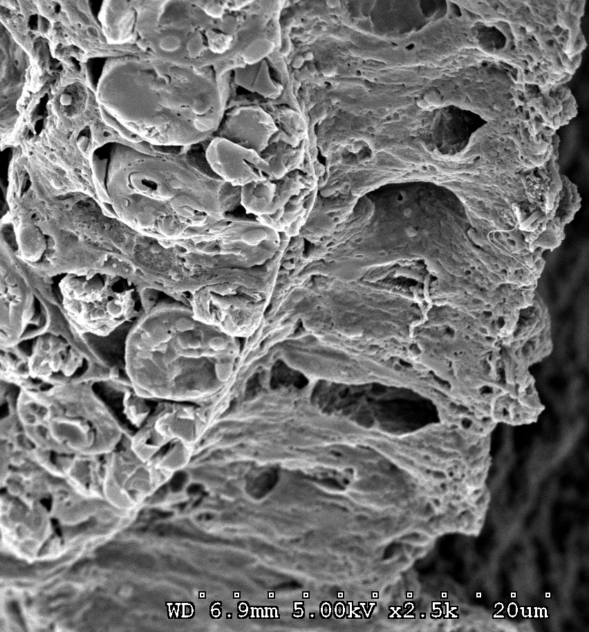
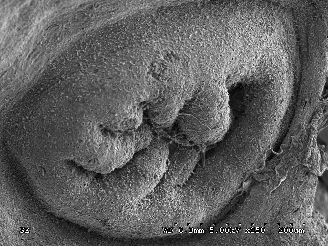
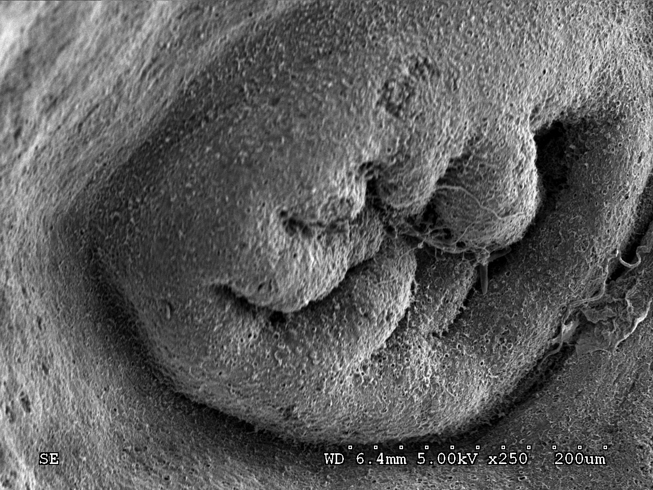
Here are all the images of high-temperature (30˚C) and control samples (27˚C) after 36 weeks of incubation.
C36T1S1 (control temperature-36 weeks-tank 1-sample 1)-freeze-fractured images only
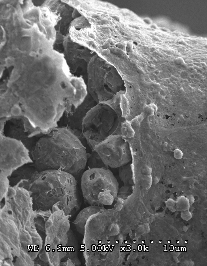
C36T2S1 (control-36 weeks-tank 2-sample 1)
freeze-fractured non-freeze-fractured
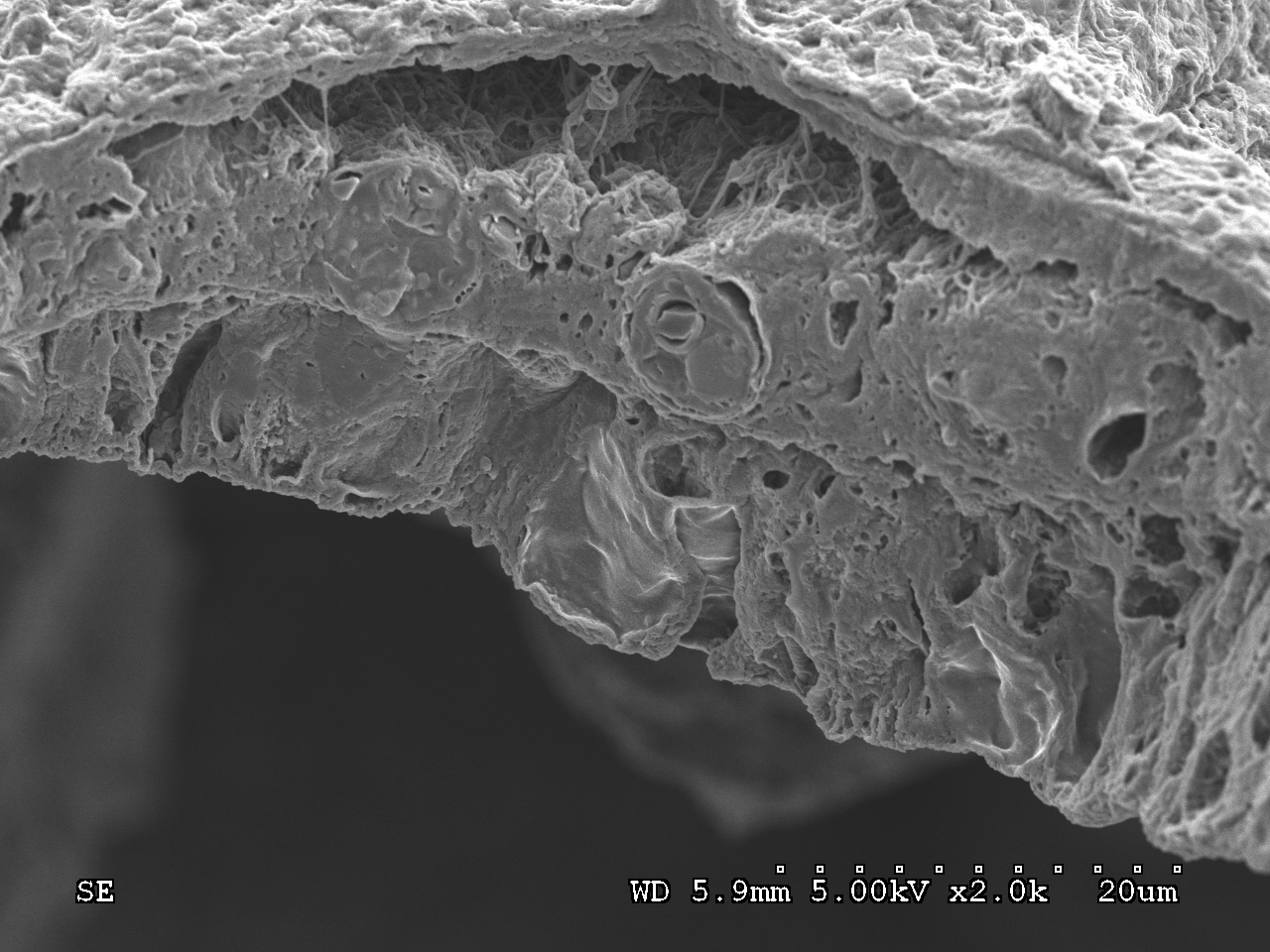
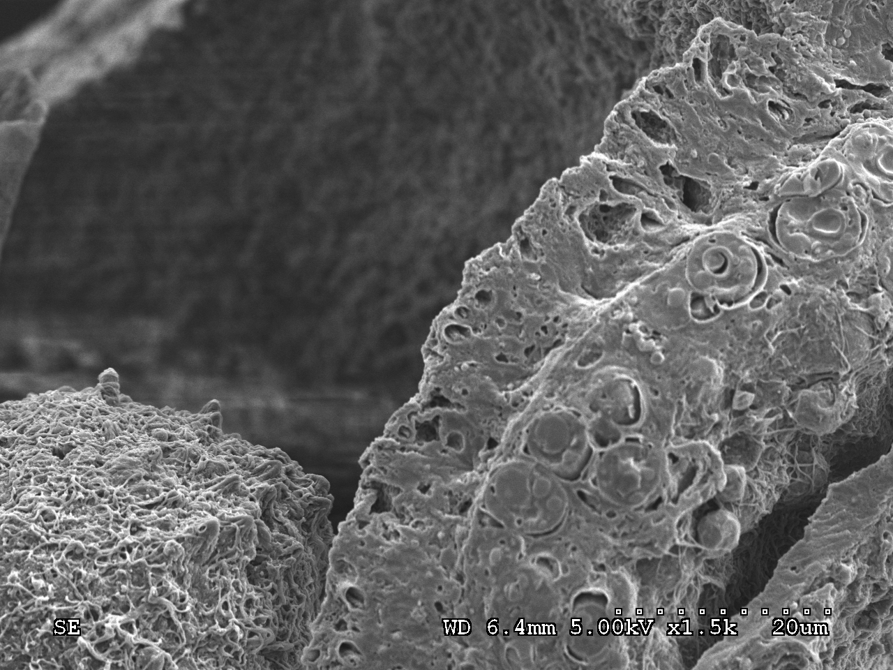
C36T3S1-freeze-fractured only
H36T1S1 (high-temperature [30˚C]-36 weeks-tank 3-sample 1
freeze-fractured non-freeze-fractured
H36T2S1-freeze-fractured only

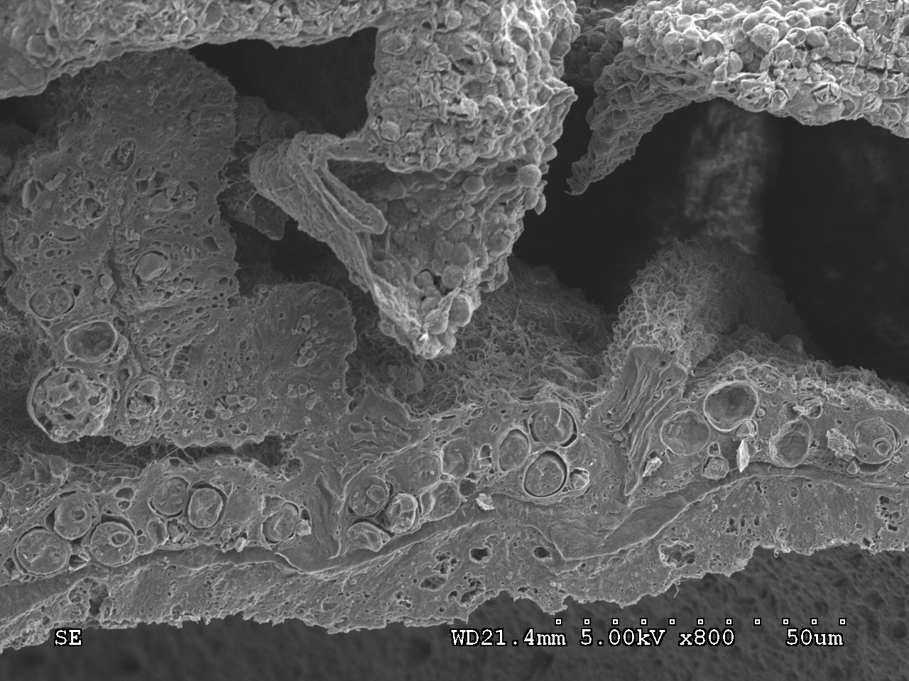
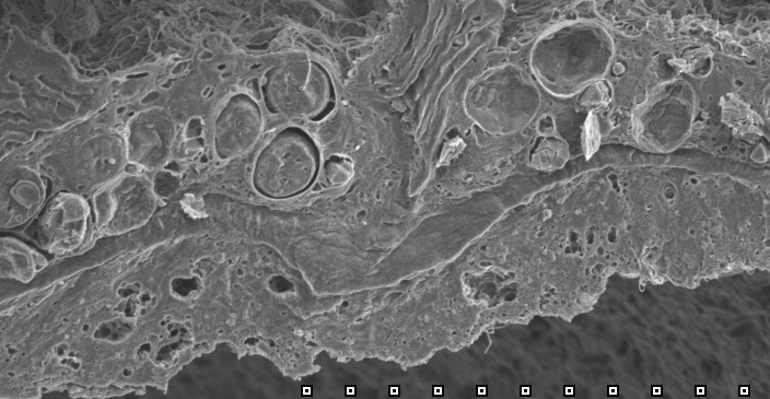
H36T3S1
non-freeze-fractured freeze-fractured